Hygroma, causes
A hygroma, which is a cavity with fluid (or a cyst), is usually classified as a benign neoplasm.
Due to the fact that the degeneration of cells characteristic of tumors is not detected in the hygroma, it cannot be called such. In accordance with the International Classification of Diseases, it is classified in the category called “other soft tissue lesions.”
The essence of the disease is this: all joints are surrounded by articular capsules, which play the role of protecting them from damage, and synovial fluid provides nutrition to the cartilage tissue. The connective tissues of the tendon sheath, called their sheaths, also have a similar structure.
For one reason or another, a person may experience protrusions on the surface of these bags or vaginas, which are filled with synovial fluid. They are subsequently separated from the vaginal cavity. This separated area becomes a capsule for the cyst, the so-called cystic formation. When an inflammatory process occurs, serous fluid begins to accumulate in it, and protein and mucus often appear in its composition.
Thus, the formation of a hygroma occurs.
— predisposition to them at the genetic level;
- prolonged load on the joint during the same type of movements (in athletes, in such working professions as seamstress, painter, plasterer);
- injuries;
— inflammation (arthritis, bursitis, etc.);
- tight shoes;
- arthrosis.
Hygroma is a complication that occurs as a result of the influence of various pathological processes occurring in the musculoskeletal system. Very often the cause of the formation of hygroma cannot be found out.
Hygroma symptoms
Prognosis and prevention
Most often, with hygroma on the hand, the prognosis is good. In some patients, the tumor becomes inflamed and then increases in size. Relapses are possible after conservative treatment (in approximately 80% of cases). After surgery, a repeat cyst appears if the excision is performed incorrectly.
Attention. Preventive measures to prevent or re-formation of cysts on the wrist should be carried out in patients at risk (office workers, professional athletes, people with a genetic predisposition, etc.).
To prevent the formation of a synovial cyst, the following rules must be followed:
- Protect hands from sports, household or work injuries.
- During training, use special orthopedic devices that fix the wrist.
- After damage to the wrist joint, you need to urgently undergo examination by a doctor.
- If inflammatory diseases of the joint appear, proper treatment must be carried out according to the doctor’s recommendations.
- You need to alternate between work and rest to avoid overloading your hands.
- If you have a hereditary predisposition, you need to choose a job where you don’t have to constantly strain your wrists or perform monotonous hand movements.
- It is recommended to do exercises daily to strengthen the joints of your hands.
Hygroma of the wrist is a common pathology, especially in patients who lead an active lifestyle or, conversely, perform monotonous work with their hands. It is important to remember that with timely treatment, the tumor is easier to treat. When its size increases, it is much more difficult to get rid of it. To achieve success in treatment, do not hesitate to visit a doctor.
Hygroma symptoms
Hygroma can be single-chamber or multi-chamber, depending on the number of formations created. They may have different structures. Sometimes the cavity has a closed shape, sometimes with the help of anastomoses it is connected to the cavity of the bursa or tendon sheath. Hygromas are of the valve type, in which fluid flow occurs only when a certain type of load is placed on the joint.
In appearance, cystic formations resemble a round lump, the size of which can range from 0.5 to 3 centimeters. It is usually mobile and has no connection with the skin. In some cases, the diameter of the cone can reach 5 centimeters. The growth rate of hygroma varies from person to person. Sometimes it grows very quickly, and sometimes it can remain constant for months or even years. The skin most often has a normal color and appearance, but sometimes there is keratinization and slight peeling.
Forecast
If this pathology is diagnosed in a timely manner, you will be prescribed appropriate treatment. In this case, the outcome of the problem is extremely favorable. You will quickly get rid of the tumor, the risk of relapse will be extremely low. In the initial stages, medications that should be regularly applied to the skin will help smooth out the tumor. Advanced hygromas can only be treated surgically.
Remember, when the first signs of the formation of this tumor appear, you need to consult your doctor as soon as possible. This will help avoid complications and allow you to quickly get rid of the problem.
Where does hygroma appear?
- on the wrist joint;
- on the knee joint;
- on the ankle joint (on the anterior lateral surface);
- on the hands and fingers;
- on the back of the feet.
It is much less common to observe hygromas on the elbow joint and in the area of the costal joints on the chest.
The symptoms of hygroma are either very weak or absent altogether. Patient complaints may only be associated with minor pain. A cystic formation causes pain only when its rapid growth begins to compress the surrounding tissues and nerve endings.
If we talk about the danger of this kind of education, then it is, in principle, absent. Not being essentially a tumor, it will not develop into a malignant formation. Sometimes, as a result of mechanical action, a hygroma can rupture, pouring its contents into the tissues surrounding it. But there is no danger in this either, since the hygroma fluid is completely sterile. The occurrence of a defect in the skin of a hygroma when it ruptures is dangerous, as it will become a gateway for infections that can lead to purulent complications.
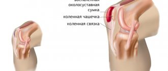
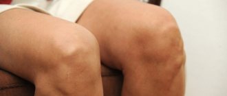
Hygroma of the knee joint
Hygroma of the knee joint is usually understood as the formation of a special cavity of connective tissue with exudative contents. It is important to know not only what hygroma is, but also how it manifests itself. This pathology can have both acute and chronic forms. At first it is completely asymptomatic. Only as the pathology progresses does the patient begin to feel pain in the kneecap area, especially during movement. The prognosis of the disease is quite favorable, complete recovery occurs in almost every case.
The main reason for the development of knee hygroma is the thinning and further protrusion of the walls of the synovial bursa.
In turn, the thinning of the walls of the bag is influenced by the following factors:
- Diseases of the joints, accompanied by infection in the cavities of the joints.
- Increased stress on the knee joint, for example, systematic lifting of weights, the need to walk a lot, professional sports.
- Any injuries to the knee joint.
- Hereditary factor.
- Diseases associated with metabolic disorders. A good example is diabetes mellitus.
Hygroma is not related to the patient’s age; it can be observed even in children. In this case, its development is usually associated with the not yet fully developed musculoskeletal system and increased vascular permeability.
hygroma of the knee joint
The risk group includes:
women (the likelihood of developing hygroma is 3 times higher than in men);
people who, due to the nature of their work activities, perform monotonous movements.
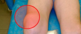
Despite its impressive and frightening size (up to 6 cm), hygroma does not cause inconvenience and, most importantly, cannot degenerate into a malignant neoplasm. It may be clearly visible on the knee. When inflammation begins, patients complain of pain in this area (increasing with flexion), limited joint mobility, and pain radiating to the ankle.
The diagnosis can be established or confirmed using ultrasound or MRI. Treatment is prescribed depending on the severity of the disease. This may be drug therapy, surgery or physical therapy.
Forms of the disease
Depending on the location of the pathological process, the following are distinguished:
- hygromas of the wrist joint of the hand - localized on the back, front or lateral surface of the wrist joint, in the area of the dorsal transverse ligament of the wrist, less often on the palmar surface of the wrist joint;
- hygromas of the back of the fingers - located in the area of the interphalangeal joint or at the base of the distal phalanx;
- hygromas of the palmar side of the fingers - are formed from the tendon sheaths of the flexor muscles of the fingers, can occupy one or two phalanges, less often develop at the base of the fingers;
- hygromas of the distal part of the palm - develop from the tendon sheaths of the flexor fingers; have a dense consistency, therefore they resemble bone or cartilage formations.
The diameter of the neoplasm, as a rule, does not exceed 3 cm, but cases of 6-centimeter hygromas have also been described.
Hygroma of the ankle joint
Hygroma of the ankle joint
This is a benign tumor that forms in the tendons and the ankle joint itself. There are no exact reasons for the formation of hygroma in this area.
Typically, the following factors contribute to its development:
Hereditary factor.
Injury to the joint or tendons in this area, including systematic and invisible microtraumas.
Inflammatory processes of the joint and tendons.
Consequences after operations.
Ankle hygroma will not go unnoticed, as it is a fast-growing tumor. It can reach the size of a chicken egg. Often the tumor grows particularly rapidly—up to several days. Despite such rapid formation, it does not pose a direct threat to the human body and does not degenerate into a malignant formation. At the same time, it is fraught with another danger: having reached a certain size, the hygroma compresses nearby blood vessels, nerves and tendons.
Usually a person begins to experience pain, limited movement, and numbness in the foot. In such cases, surgical intervention is prescribed. Sometimes the development of hygroma is asymptomatic, then drug treatment is used and its growth is simply monitored.
Hygroma can be recognized by a clearly defined contour, soft and elastic structure, free movement of the skin over the tumor, slight redness and peeling of the skin in this area.
Tendon ganglion - and a tumor is not a tumor, and bursitis is not bursitis
Have you been trying to heal your JOINTS for many years?
Head of the Institute for Joint Treatment: “You will be amazed at how easy it is to heal your joints by taking every day...
Read more "
Tendon ganglion or according to the principle of communicating vessels. At first glance at this pathology, an analogy arises with a pea or a cherry caught under intact skin, only the mobility of the “cherry” is significantly limited by the affected area.
Binding to the affected joint or tendon for a tendon ganglion (hygroma or ganglion) is mandatory, and at the same time serves as the most important differential diagnostic difference from other pathologies of the connective tissue of the extremities.
Doesn't look like a tumor...
Although the ending -oma is always a hint of a tumor, hygroma is not a tumor. After all, a tumor (especially a malignant one) is a tissue that, growing rapidly and aggressively, takes over the entire living space, crushing neighboring tissues.
This neoplasm grows extremely slowly, and then only when there is a need for its existence, under certain conditions.
The second name: tendon ganglion (ganglion - knot, hardening, compaction) also does not clarify anything - there is no trace of any special density here. To the touch, this formation of elastic-pliable consistency resembles a rubber ball not too tightly filled with air.
What then is this clearly unnecessary and unpleasant in appearance “bloating”, which so spoils the aesthetic perception of the body and significantly complicates life, especially when there is more than one such formation? This can be understood by familiarizing yourself with its structure and contents.
An autopsy shows that the ganglion is a capsule bag (cavity) with walls made of cartilage from the wall of the articular capsule, filled with pale yellow liquid with a small amount of mucus and fibrin threads.
The fluid in the cavity of the tendon ganglion is never purulent or bloody, it is always serous, and always aseptic, therefore its presence in the cavity never leads to inflammation of the tendon sheath or the wall of the joint capsule from which it is formed.
Looks like bursitis, but not bursitis
This formation is nothing more than a buffer created by the body to protect a certain area of the osteoarticular system from the load acting on it, either excessively strong, or long-term or constant.
An excessively strong load occurs during an injury (a blow to the finger with a hammer, hence the most common formation of tendon nodes on the back of the hands), and by duration we mean exposure to pressure or friction more or less constant.
This may be compression of the feet by shoes that are not removed during the day, especially tight, “tight” ones. That is why tendon ganglia also appear quite often on the back of the feet. The pressure of excess body weight on the lower extremities causes their formation in the area of the knee joints.
It is to protect against such “close” and traumatic influences that elastic “pillows” or “bubbles” are formed over the tendon and joint structures, softening and absorbing shocks and the vagaries of fate.
This pathology is very similar in appearance to bursitis, but it is not bursitis for two reasons:
- Firstly, the tendon ganglion is an artificial formation that arose from external influence. In fact, it is a hernia of the joint, a “bubble” that is “blown” out of one of its walls due to an excessive increase in pressure inside the joint. A “bubble”, most often communicating with the joint cavity through a narrow neck or anastomosis. Bursitis is a pathology of the joint capsule itself.
- Secondly, ganglion is not an inflammatory disease. Yes, its basis-wall is cartilage, as with bursitis, and this cartilage is deformed, modified. But it is not inflamed, the process of its transformation is aseptic, and not infectious or allergic in nature.
If the joint cavity is a natural formation and necessary for the body, then the ganglion cavity with its non-inflammatory contents arises from the influence of a purely external cause.
Therefore, this neoplasm should be considered a cyst, created as a result of degeneration of the synovial membrane (and filled with thickened synovial fluid).
The same banned issue for which Ernst fired Malakhov!
Joints and cartilage will be cured in 14 days with the help of ordinary...
Arising for a reason or without a reason
The mechanism of ganglion formation becomes clear if we remember that the tendon sheath, like a joint, is a closed cavity filled with fluid, which exerts a certain internal pressure on the walls of this cavity.
A sudden increase in external pressure with axial load on the joint leads to a sharp reduction in the volume of the joint cavity.
Liquid, locked in a confined space and sharply compressed, puts pressure on the walls of the cavity containing it, looking for somewhere to splash out.
And this sharp impulse, similar in force to an explosion, pushes through the wall of the articular cavity at its weakest point, causing the formation of an additional cavity - it “blows out” the hernial “bubble”, into which part of the fluid is “shot out”.
The reason for this mechanism to be triggered must be lightning fast (injury, including sports).
But more often, the basis for the occurrence of pathology is chronic injuries, in the form of a moderate, but constant or prolonged load repeated day after day.
These are movements based on the type of work performed:
- laundresses;
- cooks;
- pianists;
- seamstresses and embroiderers;
- constantly working on a computer keyboard or typewriter.
But the causes of tendon ganglion may be different:
- tight shoes, frequent formation of calluses;
- consequences of surgical interventions on joints and tendons, or their diseases leading to cartilage degeneration: arthrosis, arthritis, bursitis, tenosynovitis, or their self-medication;
- hereditarily caused weakness of connective tissue.
Often the cause of hygroma generally remains unsolved.
Structural features
Depending on the cause of its occurrence and the characteristics of the articular cartilage subjected to compression, the formation of a tendon ganglion is possible:
- single-chamber;
- multi-chamber.
In turn, the ganglion chamber, once formed, may forever lose communication with the joint cavity that gave birth to it - the anastomosis between them remains, but it is tightly closed and fluid does not move through it, an isolated type of cavity arises.
Or the anastomosis remains active, allowing synovial fluid to flow freely in both directions.
In the third option, a valve is formed in the anastomosis between the cavities, allowing fluid to pass in only one direction - into the cavity of the ganglion when the pressure in the joint cavity increases.
Clinical picture
A small tendon node does not cause pain. Problems begin as the tumor grows due to continued traumatization during work and everyday life.
In addition to the discomfort and unaesthetic appearance of this tendon-cartilaginous formation, the picture of the disease is complemented by the following symptoms:
- moderate pain of varying nature in the formation itself;
- restriction of functions (movements and sensitivity) of the involved joint or in a part of the body distal to the block that has arisen;
- deformation of the nail due to disruption of tissue trophism;
- changes that occur in the skin covering the tumor (hyperemia or cyanosis, thickening or thinning of its surface, which can lead to self-opening).
The pain can be nagging or aching, occur when pressing on the ganglion or spontaneously, when extending and flexing the blocked joint, while the skin sensations can be of the nature of “numbness” or itching.
Multiple ganglia formed on the hand or foot can generally seriously limit the range of movements in the entire limb.
There is only one solution - excise as early as possible!
Regardless of the size, which can be simply huge, and the purely technical difficulties caused, the neoplasm grows slowly or does not grow at all, it is characterized by a highly stable cellular structure and is not prone to malignant degeneration.
But at the same time:
- there is a high probability of its damage with subsequent suppuration;
- there is a tendency for further growth;
- the appearance of neurological symptoms or trophic disorders that arise in the limb from mechanical compression by the formation of vascular and nervous lines is possible;
- there is a limitation in the range of motion in the joints
This is what brings the patient to the surgeon's office. And very often this happens after persistent unsuccessful self-medication, one of the “methods” of which (crushing the tendon node) should be called simply savage.
Failure in treating a tendon ganglion by pumping out the fluid with a syringe or simply opening its cavity is easily explained by its structure: if the communication between the cavities of the joint and the formation remains, then the cavity of the ganglion will easily be filled again with fluid coming from the articular cavity.
Only radical excision of the formation with simultaneous restoration of the integrity of the walls of the articular cavity, without leaving an anastomosis, can lead to complete restoration of the functions of the affected limb or joint.
Both traditional surgical and laser excision of this neoplasm, albeit benign, are used. And you should part with it as early as possible, before it becomes a serious obstacle to work, home and life.
Surgery to remove the tendon ganglion:
To prevent relapse
To avoid the reappearance of the tendon ganglion after its surgical removal, you should strictly adhere to the recommendations of the surgeon regarding your work and lifestyle. Because premature load on a limb, despite its apparent insignificance, can easily become excessive.
With a genetic predisposition to this pathology, it is impossible to exclude the possibility of spontaneous appearance of new “nodes” in addition to existing ones.
In the same case, when we are talking about such a cause of hygroma as chronic microtrauma of joints and superficially located tendons, care should be taken when performing work and measures should be taken to change working conditions.
Hygroma of the wrist
Hygroma of the wrist
The wrist can be affected by this disease. It all starts with the formation of a small knot (about 5 mm), completely painless and not constraining movements. Usually the disease affects the joint that has experienced the greatest load.
A growth in the form of a capsule appears on the joint, with fibrous contents inside. This capsule can have one or several chambers. The first option is most common. The presence of several chambers in the hygroma indicates that the disease is in an advanced form, or complications have already begun.
Hygroma of the elbow area
Hygroma of the elbow area
Hygroma of the elbow joint can reach 5-150 mm. If at a small size it can simply not be noticed, then as it develops it becomes obvious. This may be one tumor with clear boundaries, or it may be several lumps connected to each other. In the latter case, the general contour can take on completely different shapes.
A distinctive feature of hygroma is its complete immobility, despite the fact that the skin moves calmly and even painlessly over its surface. This is explained by the fact that as the hygroma grows, it grows into the surrounding tendons, tissues and muscles.
Many patients are interested in why hygroma is dangerous. It has nothing to do with oncology; it cannot degenerate into a malignant tumor. But as the tumor grows, the hygroma still causes some discomfort to the person. This is due, first of all, to a partial loss of joint mobility and progressive pain.
If you wait until inflammation occurs, the pain becomes intense, there is numbness in the arm, and an inability to bend the arm at the elbow. In the absence of the necessary treatment, bursitis often occurs - this is a condition when inflammation also affects the fluid inside the joint.
The sooner treatment is started, the better. At first, it is quite possible to get by with taking medications. In more advanced forms, a simple operation is used that takes no more than half an hour. Immediately after it, the person can go home.
Diagnosis of hygroma and treatment of hygroma
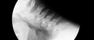
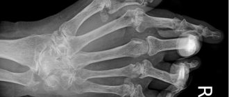
Any swelling is cause for concern. This also applies to hygroma. Despite the fact that it does not pose a danger to the body, it is necessary to differentiate it from other neoplasms. Therefore, a comprehensive diagnosis must be carried out, which includes X-rays, ultrasound, CT scans of both the hygroma itself and nearby tissues.
The most accurate diagnosis is provided by histology of the liquid contents of the formation. To do this, a puncture is taken, and the hormonal drug diprosan is injected orally. In case of infection, antibiotics are used.
Sometimes hygroma can resolve on its own.
To speed up the process, you should use:
- ointments containing diclofenac, voltaren, indomethacin;
— physiotherapeutic procedures such as ozokerite, electrophoresis, magnet, etc.;
- compresses and lotions made from infusions and decoctions of herbs, recommended by traditional medicine.
Hygroma can be destroyed with strong compression. The low effectiveness of this method, like all those described above, lies in the fact that it preserves the integrity of the capsule. It may fill with serous fluid again, and everything returns to normal. Mechanical compression is also dangerous due to fragmentation of the capsule upon rupture, which can result in the formation of small hygromas.
hygroma treatment
From all that has been said, the only correct conclusion follows: for effective treatment of hygroma, surgical intervention should be used, using surgical removal of the entire capsule using the enucleation method. Increasingly, not a mechanical incision, but a laser beam is used to remove the formation. The operation is performed under local anesthesia, and the wound formed after the procedure is covered with a sterile dressing with an aseptic preparation.
It is useful, together with this bandage, to immobilize the joint affected by the hyrgoma, using plaster or an orthosis in the form of a bandage, corset, or special shoes.
Immobilization of the damaged joint should last from 3 to 4 weeks. Throughout the entire period, it is necessary to continue treatment with anti-inflammatory and antibacterial agents; when the wound is completely healed, physiotherapeutic procedures are indicated.
Folk remedies
It will be possible to get rid of hygroma using traditional medicine methods only if the neoplasm is small and single. Otherwise, you will not achieve any result, but will lose valuable time. Many experts recommend taking a comprehensive approach to therapy and using such methods as an auxiliary treatment. The most popular remedies for hand hygroma are:
- Paraffin applications - take 100 grams of paraffin, add a few drops of essential oil, heat it all in a water bath and carry out the procedure.
- Warming compress - take a small amount of wormwood, pour 100 ml of alcohol into it. Leave the medicine to infuse for 4-6 days. After this period, use the tincture as a compress. It is best to leave it overnight.
- Bath - to prepare it, take 1 liter of water, add 1 tablespoon of soda and a few drops of iodine. When the water reaches 40-45 degrees, put your hand in it and hold it until it cools completely. It is best to carry them out 2 times a day: morning and evening.
When is surgery needed for hygroma?
Surgical removal of hygroma is indicated in the following cases:
- Rapid growth of the tumor (more than 10-15 mm), severe discomfort from it, unaesthetic appearance.
- Pain of varying intensity even at rest.
- Decreased motor activity of the joint, inability to bend the upper or lower limb, decreased performance.
- Increased likelihood of capsule rupture and spread of its contents into surrounding tissues and tendons.
- Connection of several capsules.
Surgery is possible only after a complete examination of the patient. To do this, he is assigned standard studies:
urine and blood tests (for HIV, biochemistry, STDs, hepatitis);
fluorography;
ECG.
Contraindications to surgery are exacerbations of infectious diseases, blood diseases, elevated body temperature, exacerbation of severe pathologies and pregnancy.
The operation is carried out in three ways:
1. Radical intervention involves excision of the tumor along with the membrane. After such an operation, relapse is excluded, the lump disappears immediately and does not appear again. It is performed under local anesthesia.
2. Laser removal is a modern alternative to the first option. The intervention is not only effective, but also painless, does not leave a scar, and does not affect surrounding tissues.
3. Puncture is pumping out the contents of the capsule using a syringe and then pumping into the cavity the medicine that is necessary to resolve the hygroma from the inside. After such an intervention, relapses occur.
Conservative treatment
Treatment of hygroma without surgery is indicated in cases where there is inflammation of the tissues surrounding the tumor. However, this option is only possible in the case of aseptic inflammation. Purulent inflammation is treated exclusively surgically.
Drug treatment. For therapeutic measures, drugs such as nimesil for a general effect, topical diclofenac, clemastine and histane for an antihistamine effect, as well as diprosalic are recommended. The method of application is determined by the doctor, based on the individual characteristics of the patient.
Top
Complications with hygroma
One of the most likely and dangerous complications of hygroma is its spontaneous opening. As a result, all contents enter the surrounding tissues. The capsule itself is restored and refilled over time. In this way, a multi-chamber hygroma can form - these are several neoplasms connected to each other.
In the place where the contents from the capsule enter the tissue, an inflammatory reaction appears. If you ignore this condition, tissue suppuration, infection, and even necrosis of surrounding tissues may occur.
Even if the capsule remains intact, the hygroma poses some danger. It consists of a compression effect on the surrounding blood vessels, nerves, and tendons. If no measures are taken to cure this disease for a long time, blood circulation in the limb is disrupted, immobility of the affected joint progresses, and numbness is observed. All these consequences significantly worsen the patient’s quality of life.
Diagnostic methods
The diagnosis is made on the basis of anamnestic data and characteristic symptoms. But radiography is required to exclude concomitant arthrosis, arthritis, bursitis, and synovitis. If the doctor has doubts when making a final diagnosis, then other instrumental studies are prescribed:
- Ultrasound is indicated to assess the structure of the tumor, detect fluid in it, and in the walls of blood vessels;
- MRI, CT are performed if the formation of nodes is suspected, to determine the structure of the walls and contents of the cavity.
A differentiation is made between hygroma formed in the palm area and tumors that form on cartilage and bone tissues. Also, when conducting research, lipomas, atheromas, and epithelial traumatic cysts are excluded. In some cases, using a puncture, fluid is removed from the tumor to study its contents.