Anatomical defects, also known as heart defects, are particularly common. They can be congenital or acquired during the development and aging of the body.
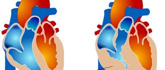
Right ventricular hypertrophy (abbreviated RVH) is the proliferation of the muscle layer of the myocardium at the level of the said chamber.
In most cases, the condition is formed under the influence of pressure overload, as well as the volume of blood entering the chamber. This is usually the result of excess blood pressure or excessive physical activity.
Thickening of the wall leads to functional impairment, but is extremely rare as a cause of death. The probability of death is determined to be 3-5% for at least 7 years or more.
Treatment as such is impossible, since the defect develops at the cellular level. However, there is a good chance of stopping the symptoms and bringing the deviation under control.
Therapy is carried out by a cardiologist; to identify the condition, a minimum list of diagnostic measures is sufficient: echocardiography, routine measurement of pressure and heart rate.
General information
Myocardial hypertrophy is understood as a clear increase in myocardial mass, which develops against the background of hypertension , pathology of the heart valve apparatus and other diseases that are accompanied by prolonged overload of a certain part of the heart.
Myocardial hypertrophy is more typical for the left ventricle, but hypertrophy of the right ventricle and atria may occur. As hypertrophy develops, the myocardium of a certain part of the heart thickens, which leads to a change in its shape, volume and size. Today, hypertrophy is considered not so much as a specific damage to the heart in arterial hypertension, but as a risk factor for the development of heart failure and sudden death.
An increase in myocardial mass is recorded in 16% of men and 19% of women under the age of 70 years. In the age group over 70 years old, these figures correspond to 33 and 49%.
The mechanism of development of the anomaly
The path of formation is determined by a violation of the normal functional activity of the heart muscle. Hypertrophy is the proliferation or increase in mass of cardiac structures in the area of the left ventricle.
Appears as a result of hypertension, less often of third-party pathologies. They have one thing in common: increasing the load on normal tissues.
Excessive stimulation, inadequate to the real capabilities of the organ, leads to the work of the adaptive mechanism. To contract more actively, the heart is forced to build muscle mass.
But this is an inherently flawed way of recovery. The bulky, expanded anatomical structure is unable to function normally.
Hence the problems with myocardial contractility, the volume of blood ejection and the functioning of cardiac structures in general, hypoxia, atrophy of tissues and systems.
Multiple organ failure occurs as a natural, deadly result.
Fortunately, the process is slow. From the moment the first symptoms of hypertrophy appear to the development of generalized disorders, it takes from 7 to 15 years.
Pathogenesis
A healthy, not organically changed heart is characterized by normal thickness of the walls of various cavities:
- left ventricle – 9-11 mm;
- right ventricle – 4-6 mm;
- left and right atrium – 2-3 mm.
Most often, hypertrophy is observed in the left ventricle, the thickness of which can reach up to 3 cm, and the mass of the entire heart can reach several kilograms. Such changes negatively affect the functioning of the entire cardiovascular system and lead to the development of heart failure .
The load on the heart can increase for various reasons, but all of them lead to thickening of the myocardium as a compensatory response to the increasing load. At the first stages, the patient does not notice any changes, but as trophism and nutrition of cardiomyocytes weaken, the vascular bed loses the ability to cover the needs of the increased myocardial area. Due to the lack of oxygen and nutrients, the contractility of the myocardium is weakened.
Just like blood vessels, the conduction system of the heart cannot expand indefinitely following the myocardium, so arrhythmias due to disturbances in impulse conduction. The thickened myocardium gradually begins to be replaced by connective tissue, losing its pumping function. Long-term hypertrophy can lead to diffuse cardiosclerosis .
Thickening of the wall of one chamber of the heart inevitably leads to expansion of other cavities if left untreated. Elimination of the causes and properly selected therapy leads to regression of LVH.
Possible consequences
If the pathology is not treated in time, it will lead to dangerous health consequences. One of the most serious complications is cor pulmonale. It is characterized by the following symptoms:
- severe and sharp pain in the chest;
- sudden drop in pressure;
- progressive enlargement of the liver;
- swelling of the veins of the neck;
- sudden psychomotor agitation;
- sudden occurrence of abnormal pulsation.
If a thromboembolism occurs, the person will experience symptoms of shock within just a couple of minutes. They are accompanied by severe pulmonary edema.
In such a situation, a large amount of transudate from the capillaries enters the lungs. This causes severe shortness of breath and chest tightness. Then suffocation, blue skin, and coughing occur. In approximately 30% of cases, the anomaly is fatal.
Classification
According to the location of the thickening:
- right ventricular hypertrophy;
- left ventricular hypertrophy;
- right atrial hypertrophy;
- left atrial hypertrophy.
Variants of left ventricular hypertrophy
The widespread use of echocardiography makes it possible to classify the architectonics of the left ventricle in hypertensive patients into 4 geometric models taking into account myocardial mass:
- Concentric hypertrophy of the LV myocardium - an increase in the relative wall thickness of more than 0.45 and an increase in myocardial mass. Symmetrical hypertrophy is formed as a result of thickening of the muscle itself, but without increasing the cavity. In certain cases, a decrease in the cavity of the left ventricle is observed. This type is most often found in arterial hypertension.
- Eccentric hypertrophy. The asymmetric form is characterized by simultaneous enlargement and thickening of the myocardium of the left ventricle and its cavity. This option occurs in cardiomyopathies , heart defects and ischemia .
- Concentric remodeling . Characterized by thickening of the wall, while maintaining normal myocardial mass.
- Normal LV geometry. The mass and wall thickness remain within normal values.
There are different degrees:
- Moderate left ventricular hypertrophy is a slight thickening of the heart cavity, which is a consequence of hypertension or other pathology of the cardiovascular system. Moderate hypertrophy signals an overload of the heart and an increased risk of myocardial infarction . The pathology is often asymptomatic and is a finding during electrocardiography.
- Severe left ventricular hypertrophy . Dystrophic changes are observed, and the mitral valve located close to the septum interferes with blood flow, which causes excessive muscle strain and significantly loads the left ventricle.
Kinds
Myocardial hypertrophy is a heterogeneous pathology. The structural features of the heart muscle allow it to increase in both length and thickness. Therefore, depending on the structure of the pathological area, the following types of GM are distinguished:
- Concentric - the myocardium hypertrophies as a result of an increase in wall thickness.
- Eccentric - the cardiac cavity expands and due to this, the size of the myocardium increases.
Concentric hypertrophy
The heart muscle changes more in thickness than in length. When viewed in cross-section, the myocardium appears wider, but there is no disturbance in end-diastolic volume. For this type of hypertrophy, coronary disorders are more typical, often contributing to the development of coronary heart disease. The occurrence of concentric hypertrophy is mainly associated with excessive blood pressure.
Eccentric hypertrophy
The increase in muscle fibers occurs more in length than in thickness. Because of this, the heart cavity also lengthens. The presented pathology is characterized by low peripheral vascular resistance, as well as increased cardiac output. Eccentric hypertrophy most often occurs due to the accumulation of a large volume of blood in the cavities.
Causes
The main causes of left ventricular hypertrophy:
- Hypertonic disease. With high blood pressure, a persistent and prolonged spasm of peripheral vessels is formed. This is why the left ventricle has to make more effort to push blood through than at normal blood pressure . This mechanism is associated with an increase in total peripheral vascular resistance, which leads to overload of the chambers of the heart. Gradually, the walls of the left ventricle thicken, which leads to rapid wear of the myocardium and the formation of heart failure.
- Cardiac ischemia. During ischemia, the heart muscle lacks oxygen. Cardiomyocytes cannot work efficiently without additional energy substrates, which leads to overload. As a compensatory mechanism, muscle tissue gradually thickens and LV myocardial hypertrophy develops. The cause of age-related cardiac hypertrophy is ischemic changes that develop over time.
- Myocardial dystrophy, cardiosclerosis . Connective tissue grows in the myocardium after inflammatory processes (post-myocardial cardiosclerosis) or after heart attacks (post-infarction cardiosclerosis). Myocardial dystrophy develops with anorexia , anemia, intoxication, infections, etching. After suffering a pathology, some cardiomyocytes lose their contractility, and the remaining cells take on the entire load. In this case, hypertrophy is also a compensatory mechanism.
- Dilated cardiomyopathy. With this pathology, an increase in the size of the heart cavities is observed due to overdistension. The left ventricle must do extra work to push blood out, which leads to the formation of hypertrophy.
- Heart defects. Disruption of the normal anatomy of the heart causes overload of the left ventricle due to increased intracavitary pressure in aortic stenosis , or due to volume overload, which is observed in aortic insufficiency. With other valve defects, hypertrophic cardiomyopathy of the left ventricle also develops over time.
- Congenital LV hypertrophy. Changes begin to form during intrauterine development and appear in the first months after the birth of the child. The reason lies in a genetic predisposition, which leads to disruption of the functioning of myocardial cells.
- Sports heart. In a person who has been involved in sports for a long time and professionally, thickening of the walls of the left ventricle is considered normal. Hypertrophy is due to the fact that the left ventricle takes on the main job of expelling a sufficient volume of blood for the whole body during training. Skeletal muscles require more blood flow during regular exercise, and as muscles grow, the amount of increase in blood flow in muscle tissue becomes constant. That is why the myocardium increases its mass, and the walls of the ventricle become stronger and thicker. For athletes, it is extremely important not to miss the moment when physiological hypertrophy can turn into pathological. This requires regular monitoring by sports medicine doctors.
- Idiopathic LV hypertrophy. If, as a result of a complete examination, it was not possible to identify the cause for the development of hypertrophy, then they speak of idiopathic hypertrophy, which most often implies a genetic predisposition.
Left atrial hypertrophy
From the left atrium, blood enters the ventricle through the mitral valve. With pathology of the valve apparatus, or more precisely with mitral valve stenosis, the atrium has to make more efforts to expel blood. If the valve does not close completely, then part of the blood returns to the atrium with reverse flow, which leads to an increase in the volume of atrial ejection. Similar changes occur in atherosclerosis and rheumatism . If the left ventricle is hypertrophied, then the muscle layer gradually increases in the left atrium.
Right atrial hypertrophy
Main reasons:
- sclerosis of lung tissue;
- obstructive bronchitis;
- bronchial asthma;
- ventricular septal defect;
- changes in the structure of the tricuspid valve;
- right ventricular hypertrophy;
- pulmonary emphysema;
- pathology of the pulmonary valve.
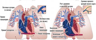
In diseases of the pulmonary system, connective tissue grows, microcirculation is disrupted and pressure in the pulmonary vessels increases. All this leads to forced hypertrophy of the right half of the heart.
Right ventricular hypertrophy, what is it?
The pathology develops after suffering diseases of the pulmonary system of an obstructive nature. Thickening of the muscle layer occurs due to increased pressure in the pulmonary circulation, which makes it difficult for the normal release of blood. Venous congestion caused by progressive heart failure can lead to right ventricular hypertrophy. Thickening of the muscle layer is also observed with congenital heart defects, with narrowing of the pulmonary valve.
Hypertrophy of the interventricular septum
Thickening of the IVS is one of the characteristic signs of hypertrophic cardiomyopathy . In pathology, thickening of the walls of both ventricles is observed with the involvement of the septum between them. This condition is only a derivative of other diseases and is characterized by a specific thickening of the myocardial walls. IVS hypertrophy is considered a fairly common pathology, observed in more than 70% of people, but most often it is completely asymptomatic.
When the interventricular septum thickens, the useful volume of the chambers of both ventricles decreases. All this leads to a decrease in the volume of blood that is released into the vascular bed when the heart contracts. The heart has to work more often to provide organs and tissues with sufficient oxygen and nutrients. Tachycardia wears out the heart muscle and leads to diseases of the cardiovascular system.
Symptoms
Myocardial dysfunction in the form of hypertrophy manifests itself with characteristic symptoms. There is some difference between lesions of the right and left sides of the heart.
Left ventricular hypertrophy is characterized by:
- discomfort in the heart area;
- rhythm disturbances;
- poor tolerance to physical activity;
- shortness of breath when walking;
- fatigue, general weakness.
Right ventricular hypertrophy is characterized by:
- blueness and paleness of the skin;
- swelling;
- heavy breathing accompanied by shortness of breath, unproductive cough;
- the appearance of arrhythmia such as extrasystole , atrial flutter or atrial fibrillation.
In some cases, autonomic symptoms, poor sleep, tinnitus, and headache .
Tests and diagnostics
At the initial visit to the doctor, complaints are collected and anamnesis is described. An objective examination includes listening to heart sounds, percussion and palpation. These methods allow you to determine the boundaries of the heart and identify its expansion. When listening to tones, the rhythm and their intensity (intensification/muffling) are assessed. Instrumental diagnostic methods are required. Indirect signs can be seen from the results of electrocardiography.
ECG. Signs of left ventricular hypertrophy on the ECG, signs of damage to other parts of the heart:
- the electrical axis is deflected to the left or located horizontally; in the V and VI chest leads, the R wave is enlarged;
- the P wave on the ECG is deformed due to changes in the atria; the “P-pulmonale” shape corresponds to the right atrium, and the “P-mitrale” shape corresponds to the left atrium;
- right ventricular hypertrophy is characterized by deviation of the electrical axis to the right, in leads V1 and V2 there is an increase in the R wave; changes in the electrical conductivity of the heart are recorded.
EchoCG. Allows you to determine the size of the heart cavities, thickening of the myocardium, calculate the pressure gradient, and calculate the mass of the myocardium during hypertrophy. Based on the results of echocardiography, it is possible to assess the pumping function of the heart and the condition of the valve apparatus.
R-graphy of the chest organs. The shape of the heart shadow is assessed; most often, the film clearly shows a hypertrophied, stretched left ventricle in the form of a characteristic protrusion at the apex.
Additionally, if indicated, coronary angiography and MSCT of the heart .
Procedures and operations
Surgical treatment is aimed at eliminating the causes that provoke the progression of LV hypertrophy. To combat ischemia, coronary angiography is performed, followed by stenting of the coronary arteries or bypass surgery (open heart surgery).
Correction of coronary blood flow allows you to normalize adequate nutrition of the heart muscle and prevent the progression of hypertrophy. In case of heart defects, prosthetics or plastic surgery of the damaged valve is performed, which also has a positive effect on the structure and function of the heart in the future. For an aneurysm of the apex of the left ventricle, excision of the muscular protrusion is performed. The operation is extremely difficult and dangerous.
Treatment with folk remedies
Traditional medicine will not help in the later stages of the disease, however, with moderate hypertrophy and minor changes, herbal therapy can alleviate the condition and reduce the risk of developing serious consequences. With treatment, it is possible to reduce shortness of breath and chest pain.
Lily of the valley in the form of gruel or tincture. To prepare the solution, pour lily of the valley with alcohol or vodka and leave in a dark place for two weeks. The tincture should be taken three times a day, 10 drops each, for two months. You can prepare a paste: pour boiling water over lily of the valley flowers and leave for 10 minutes, then drain the water and chop the softened plant. You need to take 1 tablespoon twice a day.
Herbal infusions. Horsetail, cornflower flowers, blueberries, hawthorn and motherwort improve heart function and strengthen blood vessels.
Adonis. Pour 1 teaspoon of Adonis herb into a glass of hot water and boil for 3 minutes over low heat. Leave for 20 minutes, tightly closing the lid. The resulting solution should be filtered and taken three times a day, 1 tablespoon.
Why is LVH dangerous?
In cases where minor LV hypertrophy is diagnosed in the early stages, and the underlying disease is treatable, complete cure of hypertrophy has every chance of success. However, with severe heart pathology (extensive heart attacks, widespread cardiosclerosis, heart defects), complications may develop. Such patients may experience heart attacks and strokes. Long-term hypertrophy leads to severe CHF, with swelling throughout the body up to anasarca, with complete intolerance to ordinary household stress. Patients with severe CHF cannot move around the house normally due to severe shortness of breath; they cannot tie their shoelaces or prepare food. In the later stages of CHF, the patient is unable to leave the house.
Prevention of adverse consequences is regular medical monitoring with ultrasound of the heart every six months, as well as constant use of medications.
Prevention
The development of myocardial hypertrophy can be prevented by changing lifestyle and treating the underlying disease that loads the myocardium. Key important recommendations:
- Refusal to drink alcoholic beverages.
- To give up smoking.
- Fighting extra pounds. With a weight loss of even 3-5 kg, it is possible to lower blood pressure and reduce the load on the left ventricle.
- Avoid salt and smoked foods, which slow down metabolism and promote fluid retention.
- Exercise regularly. Loads should be measured, daily. Try to walk more, Nordic walking is encouraged. Yoga and fitness classes have a positive effect on the functioning of the cardiovascular system.
Complications
In the initial stages of development, myocardial hypertrophy does not threaten the patient in any way, since cardinal changes have not yet occurred in the heart and hemodynamics are not significantly impaired. The only thing that can be observed is the clinical picture of the underlying disease - hypertension, heart disease, pulmonary disease. If the disease has entered the stage of decompensation, then symptoms of congestion in the vascular bed appear. With further progression of GM, heart failure or myocardial infarction develops.
The most severe complications are observed with left ventricular hypertrophy:
- Sudden cessation of cardiac activity.
- Coronary heart disease, which can lead to myocardial infarction.
- Arrhythmic manifestations.
Impaired functioning of the right side of the heart leads to congestion in the venous bed, which causes edema to appear and fluid to accumulate in the abdominal and thoracic cavities. In severe cases, ascites develops.
Myocardial hypertrophy in children
In pediatric practice, congenital and acquired hypertrophy of the left ventricular myocardium is considered. Acquired forms are associated with pulmonary hypertension, various carditis and heart defects. The clinical picture can be completely contradictory. Some newborn babies are too lethargic, while others, on the contrary, are too restless and noisy. Most often, during feeding and crying, the child notices blueness of the nasolabial triangle.
Older children can talk about their complaints and describe their symptoms in detail. The most common symptoms are chest pain of various types, shortness of breath even with light exertion, pallor of the skin, fatigue and lethargy. The treatment tactics for myocardial hypertrophy in children are determined by a pediatric cardiologist together with a cardiac surgeon in the presence of organic pathology.
Diet for myocardial hypertrophy
Diet for hypertension
- Efficacy: therapeutic effect after 21 days
- Timing: constantly
- Cost of products: 1600-1700 rubles. in Week
For hypertrophic thickening of the heart walls, split meals and daily intake of sufficient fluids are recommended. Avoid salt and animal fats, limit your consumption of sweets and starchy foods. Eating foods rich in omega-3 and microelements is encouraged.
Weight correction through nutrition will reduce the load on the left ventricle.
List of sources
- Shaposhnik I.I., Bogdanov D.V., “Differential diagnosis of hypertrophic cardiomyopathy and myocardial hypertrophies of secondary origin,” Breast Cancer, issue No. 12, 2014
- Chazova I. E., Ratova L. G., Boytsov S. A., Nebieridze D. V. “Diagnostics and treatment of arterial hypertension,” Systemic hypertension No. 3, 2010
- Dmitriev V. L. “Remodeling of the left ventricular myocardium in patients with stable angina according to comparison of ventriculography and echocardiography,” “From arterial hypertension to heart failure,” Proceedings of the Russian Conference of Cardiologists, Moscow, 2001
Statistics
- The disease is most often diagnosed at the age of 20-40 years.
- Men get sick twice as often as women.
- The prevalence of the disease is approximately 0.2%.
- Death from hypertrophic cardiomyopathy occurs in 2-8% of cases.
- Up to 10% of cases of GM are complicated by heart failure during a long course of the disease, the same amount is due to the development of infective endocarditis and approximately 10% to the spontaneous transition of the hypertrophic form to the dilated one.
- In the absence of appropriate therapy, death from GM is about 8%.
- Half of all deaths due to the development of GM are associated with the occurrence of more complex pathologies (complete AV block, ventricular fibrillation, myocardial infarction).