Infectious mononucleosis, caused by a virus from the herpesvirus family, is experienced by slightly more than half of people in childhood and adolescence. Statistics say: nine out of ten adults have Epstein-Barr virus DNA in their blood; apparently, many people suffer from infectious mononucleosis in a latent form.
Our expert in this field:
Nagibina Margarita Vasilievna
Infectious disease doctor, MD
Call the doctor
Why was infectious mononucleosis previously called Filatov's disease?
More than 130 years ago, children’s doctor Filatov thought of combining the symptoms of inflammation of the lymphoid organs into one disease, “idiopathic inflammation of the cervical glands,” and 4 years after that, the German Pfeiffer called the same combination of symptoms “glandular fever.”
A quarter of a century later, a completely “new” disease, infectious mononucleosis, was discovered, and a little later its discoverers realized that it had already been described under other names. In the USSR, they preferred to call the infection Filatov’s disease, emphasizing priority, but later they joined the international nomenclature.
When can you get vaccinated?
A disease is a disease, and vaccinations are vaccinations. It is impossible to allow a child’s body, having suffered from mononucleosis, to be defenseless against other infections.
The Internet, in search of an answer to the question of when a baby can be vaccinated (for example, manta rays), refers to the official document - guidelines 3.3.1.1095-02 on the procedure for preventive vaccinations.
According to the instructions, if the disease is mild, vaccination is possible after the patient’s condition has normalized.
If the disease is severe - after 2-4 weeks. In practice, pediatricians, as a rule, extend the period to 6-12 months. It all depends on the individual characteristics of the body, its readiness for certain tests.
Infectious mononucleosis in adults
Transmitted by a sick person or a healthy virus carrier, the Epstein-Barr virus infects immune cells - B lymphocytes and lymphoid organs: tonsils, lymph nodes, spleen and liver. On the surface of their cells there are special receptors that the virus needs to invade.
The disease is accompanied by symptoms of intoxication, and in most cases ends after a month or two with complete recovery, which, however, cannot be felt soon, since weakness and malaise continue to bother you for a long time.
If an adult becomes infected with infectious mononucleosis, the disease is more severe than in childhood, or milder and even with virtually no clinical manifestations. Atypical forms often occur, especially with age-related decreases in immunity, for example, this is typical for HIV-infected people.
Immune disorders as a result of infection can be superimposed on an age-related decrease in defenses, which is manifested by prolonged fever and intoxication in the absence of characteristic symptoms.
An individual approach is applied to each patient based on clinical recommendations from the world's leading medical centers, which allows for optimal results to be achieved in a short time.
The virus damages lymphocytes that are found in the lymph nodes, tonsils, spleen and liver, so all the symptoms of infectious mononucleosis develop in these organs, and the temperature reflects the severity of inflammation.
A disease with clinical manifestations of no more than 3 months is designated as an acute infection, if the disease lasts up to six months - protracted infectious mononucleosis, more than six months - chronic. If, after the disappearance of all manifestations, clinical signs of the disease reappear almost after recovery a month later, then this is a relapse and the disease will most likely take a chronic course.
Epidemiology
The source of infection is a sick person. The pathogen is transmitted by airborne droplets, but the disease is low-contagious. It is believed that transmission of the pathogen occurs only through close contact. There are spring and autumn rises in incidence. Mostly children and young people are affected; in people over 35-40 years of age, M. and. is very rare. The disease is registered mainly in urban residents. Ch. are observed. arr. sporadic cases, familial and group outbreaks are possible. However, wedge, and hematol, examination of persons in contact with the patient allows us to identify among them patients with mild, erased and subclinical forms of the disease. M. and. recorded in various climatic and geographical zones of the world.
Pathogenesis
little studied. Epidemiological and clinical data suggest that the entry gates of the pathogen are the pharynx and nasopharynx. Enlargement of the cervical lymph nodes from the first days of the disease, as well as reports of experimental infection of monkeys with an emulsion from cervical lymph nodes and blood taken from patients in the acute period of the disease, indicate lymphogenous and hematogenous ways of spreading the pathogen in the patient’s body. On the tropism of the pathogen M. and. systemic lymphadenopathy, enlargement of the liver and spleen, which is an expression of generalized hyperplasia of lymphoid and reticular tissue under the influence of a pathogen, also determines the appearance of atypical mononuclear cells in the blood. Almost constant hyperplasia of the tonsils and lymphoid formations of the pharynx, especially the nasopharynx, can also be considered as a particular expression of the systemic reaction to the pathogen of the entire lymphoid tissue. The influence of the pathogen on the tonsils and changes in the body's reactivity during the course of the disease often determine, with the participation of the bacterial flora of the oral cavity, the development of sore throat with overlays on the tonsils and a peculiar course. Analysis of hematological parameters does not give grounds to consider angina as a consequence of the “defenselessness” of the body due to granulocytopenia and secondary bacterial infection, since at the height of the disease there is no sharp decrease in the absolute number of granulocytes in the blood and there is no therapeutic effect from antibacterial therapy. Mononucleosis tonsillitis and a number of other wedge symptoms indicate the possibility of allergic mechanisms being involved in the pathogenesis of the disease. There are five phases in the development of M. and.: infection; lymphogenous introduction of the pathogen into regional lymph nodes and their response (hyperplasia); viremia with generalization of the pathogen in the body and a systemic reaction of lymphoid tissue; infectious-allergic phase, one of the manifestations of the cut is the undulation of the course of the disease; recovery phase, accompanied by the mobilization of immune mechanisms. Hyperplasia of lymphoid tissue is associated with hypergammaglobulinemia, increased titers of heterophilic antibodies and the level of immunoglobulins, in particular IgM. Involvement in patol, the process of the liver leads to its constantly observed functions and disturbances.
What are the symptoms of infectious mononucleosis?
The incubation period from the introduction of the Epstein-Barr virus to the development of the disease is one to one and a half months, during which there are no manifestations of infection.
Incubation ends with the beginning of the prodromal period, when the patient feels manifestations of viral intoxication: weakness and fatigue, muscle pain. At this stage there are no characteristic signs; all manifestations are inherent in any viral infection. This continues for one or two weeks.
The first signs of infectious mononucleosis occur acutely with fever, sore throat and swollen lymph nodes. All manifestations appear gradually over a week, last for a total of two weeks to a month and regress. Malaise may persist for several months after all manifestations of the infection have disappeared.
If a disease occurs, you must immediately contact an infectious disease doctor, who provides consultations seven days a week and seven days a week at the Medicine 24/7 clinic. Early initiation of therapy will avoid irreparable consequences.
How long does the rash last
The disease lasts about two weeks, then there is a gradual improvement, but sometimes the rash goes away after 3-5 days. If the treatment is carried out correctly, it disappears quickly and without complications: the spots first fade and then become completely invisible.
The rashes disappear as the main symptoms of the disease weaken. Even after no visible symptoms of the disease remain, lethargy, apathy, and weakness persist for a long time.
To speed up healing, it is necessary to normalize the immune system and metabolism, and adhere to a diet. Treatment is similar to that of a common cold. Bed rest and full sleep are required. Rest is especially important because the spleen usually increases greatly in size and needs to be protected. In severe cases, hormonal anti-inflammatory drugs may be prescribed to treat the rash to relieve symptoms.
Although mononucleosis is a highly contagious disease, the rash itself is not spread from person to person. The appearance of mononucleosis in a group of children should not become a reason for quarantine, since, despite its high contagiousness, this disease does not cause epidemics.
Thus, a rash with mononucleosis can be a symptom of the disease or a consequence of taking antibiotics. Despite the fact that skin manifestations do not require specific treatment, the patient should be under the supervision of a doctor, since specific symptoms (high temperature, changes in blood composition, damage to the oral cavity, changes in the spleen and liver) require careful monitoring.
What symptoms appear in the first week?
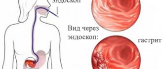
Inflammation of the tonsils develops in the first days and lasts for two weeks, and can vary in severity - from catarrhal to ulcerative necrotizing tonsillitis. A characteristic symptom is a significant enlargement of the tonsils, a bright red mucous membrane of the soft palate with a uvula, and millet-like enlarged follicles are visible on the back wall of the pharynx.
The second characteristic symptom is that the lymph nodes on the neck and back of the head symmetrically enlarge, but in weakened adults with infectious mononucleosis, all peripheral lymph nodes may enlarge, and in severe cases, internal lymph nodes as well. They are dense and painful when touched.
Every tenth person develops a rash at the end of the first week; this symptom does not have any special differences; the rash can last about a week, fade and peel off. In half of the cases, there is a connection between the occurrence of skin symptoms in infectious mononucleosis and treatment with certain antibiotics.
Only a qualified specialist with extensive diagnostic capabilities is able to quickly make a differential diagnosis and identify the cause of the pathological condition, and all this is possible in diagnostic medicine.
Treatment
No specific treatment has been found. Antibiotics and sulfonamide drugs do not have a noticeable effect on the course of infectious mononucleosis, and do not prevent the development of severe sore throat. Bed rest, diet, desensitizing agents, vitamins are indicated, and for severe sore throat - rinsing the throat with antiseptic solutions. Indications for the use of antibiotics are relative - only for the purpose of preventing secondary infection in the presence of absolute granulocytopenia and widespread lesions of the pharynx, especially in young children. There are isolated reports of treatment with DNAase and metronidazole. Indications for the use of steroid drugs are hepatitis with the presence of severe intoxication, severe lymphadenopathy and a pronounced allergic component with compression and swelling in the neck and severe hyperplasia of the tonsils, making breathing difficult, rare severe neurological or hemorrhagic syndromes. Prednisolone is prescribed in a short course, no more than 7-8 days (1.5 mg per 1 kg of weight) in the morning.
Clinical picture in the second or third week of infectious mononucleosis
In half of the patients, the spleen enlarges, usually from the second week of infection, and lasts until the end of the disease. At the same time, the liver enlarges and its functions suffer. In biochemical analysis, liver enzymes increase several times. A disorder of bilirubin metabolism is manifested by jaundice and itchy skin, but this is a rare and quickly passing symptom. Mainly concerned about weakness and indigestion, with lack of appetite. An enlarged spleen and liver is called hepatosplenomegaly.
What “wrong” variants of the course of the disease are possible in adults?
In patients weakened by chronic diseases and elderly patients, the visceral form may develop, when the virus affects internal organs, including the heart and kidneys, as well as the nervous system. In the worst case scenario, multiple organ failure develops.
But still, an erased form is more often possible, when infectious mononucleosis occurs under the guise of a mild viral respiratory infection or with no symptoms at all - an asymptomatic option, when only tests indicate the disease, and the person does not have any clinical manifestations.
For infectious diseases, the Medicine 24/7 clinic uses the most effective domestic and foreign treatment methods, which can significantly improve the quality of life of our patients, maintain their activity and prevent the process from becoming chronic. Seek help from an infectious disease specialist, call: +7 (495) 230-00-01
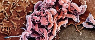
The cause of infectious mononucleosis is a virus from a small but harmful group of herpes viruses. Its number 4 out of eight types and its proper name is Epstein-Barr virus.
During reproduction on its surface, the viral agent manages to synthesize more than 70 different proteins. Different protein components are produced at different periods of life; today they have even learned to use them to determine the period of life of a pathogenic agent, and with it the stage of the disease. All these products are synthesized for a reason, but to facilitate penetration, improve survival and reproduction, and of course, to protect against the host itself.
Take care of yourself, book a consultation now
How does the infectious mononucleosis virus enter cells?
The cause of the development of infectious mononucleosis is the penetration of the pathogen into the mucous membrane of the upper respiratory tract. Mucosal cells and B-lymphocytes have a special receptor, and the virus attaches to it. It also clings to white blood cells.
Special immune cells - cytotoxic - swallow B-lymphocytes affected by the virus and digest them, releasing many biologically active substances that cause fever and intoxication in the infected patient. There is an assumption that it is these substances that cause liver damage, forming the clinical symptoms of infectious mononucleosis.
The medical center uses high-precision equipment to conduct round-the-clock examinations; quick and error-free diagnosis allows you to begin treatment immediately, including on holidays and weekends.
Causes of the disease and routes of infection
The causative agent of the disease is herpes virus type 5. The disease is caused by the activation of a pathogenic virus, which, moving through nerve tissues, causes mononucleosis syndrome. The main reason for the development of cytomegalovirus mononucleosis is the transition of the virus from a latent state to an active state when the body’s protective properties are weakened. The main causes of mononucleosis caused by cytomegalovirus include:
- sexual intercourse with a carrier of the virus;
- skin and venereal diseases;
- complications after influenza, ARVI;
- severe hypothermia of the body;
- psychological stress.
Routes of infection
The main routes of infection are:
- direct contact - kissing, sexual intercourse;
- eating from the same cutlery;
- blood transfusion;
- in utero;
- breast milk during lactation.
Why do relapses of infectious mononucleosis develop?
The intracellular residence of a pathogenic particle protects it from the effects of the immune system, which causes the formation of a chronic infection with constant carriage. There are few B-lymphocytes populated by particles, but at the slightest suppression of the immune system, they immediately begin to multiply. The cause of relapse of the disease is the division of viral DNA with the formation of new chains and the subsequent formation of new viral particles, which leads to the death of the host cell. And then the whole process goes in a new circle.
Only a qualified infectious disease doctor with extensive diagnostic capabilities can quickly make a differential diagnosis and identify the cause of the pathological condition, and all this is possible at the Medicine 24/7 clinic.
Is it possible to swim in the sea and sunbathe in the sun?
Several years ago, both adults and children after mononucleosis were categorically not allowed to swim in the sea or sunbathe.
The argument is the negative impact of ultraviolet radiation on the body’s immune barrier: it should be restored after an illness, and the sun supposedly interferes with this process.
Considering the herpes-like nature of mononucleosis and the fact that its “closest relative,” herpes type 6, under the influence of hot rays can provoke a relapse of the disease, doctors decided to play it safe in the case of mononucleosis.
Today, however, these arguments, having not received scientific confirmation, are called into question.
Evgeny Komarovsky, for example, states: you can safely go to the sea, get a tan, and swim, under one condition - observing a sense of proportion.
By the way, moderate sun exposure protects the body from the development of multiple sclerosis, which often develops in those who have had mononucleosis.
How dangerous is the chronic form of infectious mononucleosis?
Epstein-Barr is the cause of the development of every hundredth malignant lymphoma, because two of its strains living inside B-lymphocytes reprogram blood cells for malignant transformation. Like all its herpetic counterparts, the virus lives in a person for life; it configures blood B-lymphocytes for uncontrolled reproduction, turning off the program of natural cell death after completion of the normal life cycle. With a decrease in immunity, already realized by T-lymphocytes, in which Epstein-Barr is also involved, obstacles to cancerous degeneration are eliminated.
Acute infectious mononucleosis resolves in most people. To prevent the disease from becoming chronic, the help of a highly qualified infectious disease specialist who knows the clinical problem is necessary. The Medicine 24/7 clinic has the necessary equipment and, most importantly, doctors of all specialties who are interested in the recovery of patients. Get a consultation with a doctor at the Moscow Infectious Diseases Clinic, make an appointment by calling +7 (495) 230-00-01
Infectious mononucleosis has a clear and specific picture, but HIV, toxoplasmosis and cytomegalovirus infection can masquerade as it, so laboratory diagnosis is mandatory. By detecting antibodies to the Epstein-Barr virus, the course of the acute process is monitored, a chronic disease and a latent infection are identified.
How else is infectious mononucleosis diagnosed?
When a pathogenic agent is introduced into a lymphocyte, it begins to produce many nonspecific antibodies, synthesizing everything it can, including rheumatoid factor and cold immunoglobulin. These products, called heterophilic Paul-Bunnell antibodies, are also involved in diagnosis; they will completely disappear in six months or less.
When their level increases to 1: 224, infectious mononucleosis is confirmed in half of the patients in the first two weeks of the disease; by the end of the month they are detected in almost all. If they were not found during the first examination, the examination is repeated a week later.
Medicine 24/7 specialists will conduct an examination, identify the main cause of the pathology in the shortest possible time and make the correct diagnosis.
Diagnostics
To make an appropriate diagnosis, it is necessary to undergo a series of medical examinations. They include:
- Taking medical tests. To do this, you need to take a general blood and urine test;
- Examination of the patient;
- Research on herpes viruses.
Based on the test results, the doctor selects the appropriate treatment.
What “diagnostic substances” does the infectious mononucleosis virus produce?
After introduction, the viral particle produces the substance it needs for reproduction - early antigen (EA), to which antibodies are generated in the human body in the form of immunoglobulins M (IgM) and G (IgG).
Next, the virus begins to produce unique antigens from pieces of its genome. They trigger the production of special antibodies in the sick body, which are detected during laboratory diagnostics and the course of the disease is monitored based on their level. These are antibodies to the VCA viral capsule, its MA membrane, and the EBNA core.
If antibodies to VCA are absent, and the clinical manifestations are completely consistent with mononucleosis, then the infection is caused by toxoplasma or cytomegalovirus. All doubts are rechecked after a week; if they do not dissipate, then the reaction is considered negative.
Specialists always take into account not only the individual characteristics of the disease, but the capabilities and interests of each patient, so that diagnosis and treatment are as comfortable as possible.
Diagnosis
The diagnosis is based on the wedge, the picture and the characteristic blood picture.
With infectious mononucleosis, there is an increase in the titer of heterophilic antibodies, which is established using the Paul-Bunnell reaction - agglutination of sheep erythrocytes (see Paul-Bunnell reaction), Paul-Bunnell-Davidson reaction, Lovrik reaction - agglutination of sheep erythrocytes treated with papain, Tomczyk reaction - agglutination of bovine erythrocytes, Hoffa-Bauer reaction - agglutination of fresh or formalized horse erythrocytes, etc. The simplest of them and quite reliable is the Hoffa-Bauer reaction; it is positive in 90% of patients with M. and. and rarely (up to 3% of cases) in other diseases.
In the differential diagnosis, one should keep in mind diphtheria of the pharynx and nasopharynx (see Diphtheria), as well as various forms of tonsillitis (see), anginal-bubonic form of tularemia (see), diseases of the hematopoietic system (acute leukemia, agranulocytosis, to a lesser extent lymphogranulomatosis ), leukemoid reactions (see).
In the absence of angina M. and. should be differentiated from typhoid fever (see), viral hepatitis (see Viral hepatitis), mumps (see Epidemic mumps), tularemia, cat scratch disease (see Felinosis). Clinical and hematological symptoms similar to M. and. can be observed with cytomegaly (see), toxoplasmosis (see), as well as with allergic conditions.
What should you do if someone in the family gets infectious mononucleosis?
- Separate the patient from other family members.
- When interacting with a sick person, wear a protective mask, change it every 2 hours, and wash your hands up to the elbows.
- The patient is prescribed strict bed rest for a week. Provide personal utensils and everything that is required for care, preferably disposable.
- Treat all external mucous membranes with antiseptics at least three times a day to prevent bacteria and fungi from joining the inflammation.
The infectious disease specialist at the Medicine 24/7 clinic, having a broad clinical horizon, has at his disposal the most modern diagnostic equipment, performs complex tests, which affects the patient’s favorable prognosis. Sign up for a consultation by phone
What complications occur with infectious mononucleosis?
The most common is the addition of bacterial flora, when, for example, a viral infection of the pharyngeal tonsils flows into a bacterial purulent-necrotic sore throat.
Complications from the organs are quite rare; it can be damage to the nervous system in the form of meningitis. As a rule, this is possible in elderly and weakened patients in the first week of illness. Neurological manifestations may be the only symptom of the disease; in most cases, they disappear without a trace.
In very rare cases, it is possible to develop hemolytic anemia, a critical decrease in the number of leukocytes - neutropenia, thrombocytopenia, probably due to damage to the bone marrow by the virus.
Infectious disease specialists at the Medicine 24/7 clinic, during a comprehensive examination, identify risk factors and objective causes that can lead to the development of adverse complications, which makes it possible to begin preventive measures.
Pathological anatomy
The object of pathological research is Ch. arr. biopsy material of lymph nodes, removed tonsils, material obtained from a puncture biopsy of the liver. Fatalities are rare. The main causes of death are spontaneous rupture of the spleen, damage to the nervous system (meningoencephalitis, polyneuritis), secondary inf. complications, in some cases, suffocation due to extensive swelling of the larynx. At autopsy, a generalized enlargement of lymph nodes, spleen, liver, tonsils, expressed in varying degrees, and hyperplasia of group (Peyer's) patches and solitary lymphoid follicles of the colon are observed macroscopically. tract with focal necrosis, sometimes hemorrhages in the serous and mucous membranes. The pia mater and brain substance are usually plethoric.
Microscopic examination of the lymph nodes reveals significant hyperplasia of lymphoid tissue in the cortex and medulla. They are characterized by the presence of a large number of immunoblasts - blastotransformed lymphocytes (see Blastotransformation of lymphocytes) - large cells with light nuclei and a wide zone of basophilic cytoplasm, intensely pyroninophilic when stained using the Brachet method. The nuclei of these cells have a clear membrane and contain nucleoli. Among the immunoblasts there are large Hodgkin-type cells, as well as binuclear and multinuclear elements, morphologically very similar to Berezovsky-Sternberg cells in lymphogranulomatosis (see). There is hyperplasia of lymphoid follicles, sometimes an increase in the number of plasma cells and eosinophils, and there are often focal necrosis. Partial erasure of the pattern is possible due to the fusion of hyperplastic follicles in certain areas, accumulation of immunoblasts and small lymphocytes in the lumens of the sinuses, and their infiltration of interlobular septa and capsules. Severe hyperplasia of lymphocytes with the presence of a large number of blastotransformed elements is also detected in the tonsils, spleen, thymus gland, and focally in the bone marrow. Multiple nested infiltrates from the same cells are found along the connective tissue layers in the lungs, liver, myocardium and other organs. Discomplexation and dystrophic changes in hepatocytes are observed in the liver. Significant changes in the central and peripheral nervous system have been described - perivascular accumulations of lymphocytes, focal hemorrhages in the substance of the brain and spinal cord, degeneration of ganglion cells. Changes can be localized in the roots of the spinal nerves and spinal ganglia. Gistol, the picture should be assessed necessarily taking into account the wedge, data, in particular changes in the blood picture, and dynamic observation (with a repeated biopsy in the lymph node during the recovery period, the normal gistol structure is restored).