From this article you will learn:
- how to understand that you have sinusitis (signs),
- in what cases does it occur without fever,
- how to treat sinusitis in adults,
- what medications to take for sinusitis, drops, etc.
Sinusitis is an inflammation of the mucous membrane of the maxillary sinuses, which can occur as a complication during the development of acute infectious diseases (ARVI, influenza), with chronic inflammatory diseases of the nose, as well as in the presence of inflammatory foci at the apexes of the roots of the lateral teeth of the upper jaw. The presence of allergic rhinitis in a patient may also be a predisposing factor for the development of this disease.
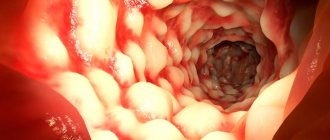
The maxillary sinuses are air-filled cavities that are lined from the inside with mucous membrane (Fig. 1-3). They belong to the paranasal sinuses and are also called the maxillary sinuses. Accordingly, sinusitis can also be referred to as “maxillary sinusitis.” Because Each person has 2 maxillary sinuses located in the thickness of the upper jaw (one on each side) - sinusitis can be unilateral or bilateral.
Maxillary sinuses on the diagram and CT images –
If you suspect that you have developed sinusitis, symptoms and treatment in adults can be very variable and depend on the form of the inflammatory process and the nature of its course. The first signs of sinusitis in adults will also be greatly influenced by the source of sinus infection. Infection can be of a rhinogenic nature (from the side of the nasal cavity), odontogenic nature (from the apex of the roots of the 5-6-7 upper teeth).
However, the most severe forms of sinusitis occur when the patient’s infection penetrates into the sinus simultaneously from both the nose and the teeth, because in this case, a mixed microflora is present in the sinus, resistant to most antibiotics. Also, a severe course is observed in patients with a long history of self-medication (regular use of antibiotics), and in such patients it can be very difficult to relieve purulent inflammation, plus they can still have a lot of complications.
Signs of sinusitis in adults -
Signs of sinusitis depend on the form of the disease - it can be acute or chronic, as well as on the nature of the inflammatory process. According to the form of inflammation, acute sinusitis is divided into serous and purulent, and chronic sinusitis is divided into catarrhal, purulent, polypous and purulent-polypous. In addition, the first symptoms of sinusitis will differ depending on what was the source of the infection - the nasal passages, or a “dental infection” associated with inflammation at the roots of the upper lateral teeth.
Complications
Sometimes with acute, but more often with chronic. G., in both adults and children, intracranial complications occur in the form of edema of the meninges, serous or purulent meningitis (see), meningoencephalitis, phlebitis of the dural sinuses with the development of rhinogenic sepsis (see), pachymeningitis (see), rhinogenic brain abscess (see Brain, abscess), rhinogenic arachnoiditis (see). They are most common during an influenza epidemic. There may be orbital complications: reactive swelling of the tissue of the orbit and eyelids, retrobulbar abscess, osteoporosis of the orbit, phlegmon, thrombosis of the veins of the orbit, etc. Periostitis of the upper jaw is also found.
Symptoms of acute sinusitis -
Sinusitis of rhinogenic origin (it is also sometimes called the term “rhinosinusitis”) - usually occurs either against the background of an acute runny nose during infectious diseases such as ARVI and influenza, against the background of allergic rhinitis, or against the background of chronic inflammatory diseases of the nose. The latter are usually accompanied by constant nasal congestion and a mild runny nose. The development of sinusitis in this case is associated with the development of swelling of the mucous membrane of the nasal passages and an increase in mucus production.
The fact is that each maxillary sinus communicates with the nasal passages through a small anastomosis (opening). These openings are designed for ventilation and physiological cleansing of the sinuses from mucus, desquamated epithelial cells that enter the sinuses of pathogenic bacteria. The resulting swelling of the mucous membrane of the nasal passages leads to an increase in its volume, which leads to partial or complete closure of these anastomoses and disruption of the outflow of mucus. This creates good conditions for bacteria to multiply and inflammation to develop.
In case of acute rhinogenic sinusitis, the patient (in addition to a runny nose and nasal congestion) is usually only bothered by malaise at first. In the initial period, the inflammation is serous in nature and the formation of pus in the sinus does not yet occur. Mucous discharge from the nose during this period still has a transparent color, without an unpleasant odor. The temperature is usually normal, or the temperature may be elevated, but this is not due to the development of serous sinusitis, but to the underlying disease (for example, ARVI and influenza). In the absence of timely measures at this stage, serous sinusitis turns into purulent.
The first symptoms of sinusitis of odontogenic origin:
According to statistics, the rhinogenic origin of sinusitis is observed in approximately 62% of patients, and in more than 30% of all cases, sinusitis is of odontogenic nature (i.e., associated primarily with inflammatory foci at the apexes of the roots of the 5-6-7 upper teeth). Although it should be noted that a number of patients may also experience a combined type of sinus infection. With sinusitis of odontogenic nature, patients may remember that the development of symptoms was preceded by pain or discomfort in one of the lateral teeth of the upper jaw (24stoma.ru).
The pain can be acute or occur only when biting on a tooth. In some patients, toothache may be absent altogether, and in this case the causative tooth can only be determined by an x-ray. Among the very first symptoms (besides toothache) there is usually only malaise. Thus, runny nose and nasal congestion with the development of sinusitis of odontogenic nature are usually absent (nasal discharge appears only later, and is not associated with a runny nose).
Inflammation at the root apex with sinusitis -
Symptoms during the transition of serous sinusitis to purulent -
With serous sinusitis, a sharp swelling of the mucous membrane of the maxillary sinus develops, which can lead to narrowing or complete closure of the anastomosis between the sinus and the nasal cavity. In addition, there is a sharp increase in mucus production, and the formation of serous inflammatory exudate (there is still no pus in the sinus at this stage). All this leads to the transition of serous inflammation to purulent, and a sharp worsening of the symptoms of sinusitis.
Acute purulent sinusitis has characteristic symptoms, which already make it possible to establish the correct diagnosis without any problems. And besides, with acute purulent sinusitis, there are already specific changes on radiographs and computed tomography (CT) images that make it possible to confirm the diagnosis.
Signs of acute purulent sinusitis:
- General symptoms: weakness, lethargy, weakened sense of smell, chills, loss of appetite.
- Body temperature rises to 37.5 - 39.0 degrees (sometimes higher). The degree of temperature rise depends on the characteristics of the immune system of a particular person’s body’s response to infection. Those. If you usually have colds without a significant rise in temperature, then you should not expect a strong rise in temperature in case of acute purulent sinusitis.
- If inflammation develops in only one sinus, a feeling of heaviness may appear in the corresponding half of the face.
- When pressing on the skin in the projection of the anterior wall of the sinus, pain may be felt. In addition, pain or discomfort may appear when tapping on the teeth located in the projection of the inflamed sinus (usually the 5-6-7 teeth of the upper jaw).
- As a rule, purulent exudate will be released from the nasal passage on the side of the inflamed maxillary sinus (its discharge increases when the head is tilted forward). In the morning, patients often notice traces of pus leaking from the nose on the pillowcase. But! If the outflow of purulent exudate from the sinus into the nasal cavity is completely disrupted, then this symptom may be absent.
- If the outflow of purulent contents from the sinus is disrupted, first periodic and then constant increasing pain may occur, which will first be localized in the projection of the inflamed sinus. Subsequently, the pain may begin to spread to the frontal, temporal, occipital regions, as well as to the area of the teeth of the upper jaw (from the side of the affected sinus).
- In severe cases of purulent sinusitis, swelling of the soft tissues of the face and their redness may also be observed on the side of the affected sinus, plus purulent periostitis of the jaw (flux) may develop.
Computed tomography (CT) and radiography –
The diagnosis of “Acute purulent sinusitis” is made on the basis of symptoms, as well as CT or radiography data. A traditional x-ray will only show the “darkening” of the sinus and the level of fluid in it (the level of pus), without any details. In turn, a CT scan will allow not only to obtain a much clearer image of the sinuses, but also to simultaneously evaluate the quality of filling the root canals of the teeth in the sinus projection, as well as see the presence of inflammatory foci at the apexes of the roots. This is very important for identifying the source of infection and choosing the right treatment strategy.
Purulent sinusitis on x-ray and CT scan –
Diagnostics
For a competent specialist, establishing a diagnosis for sinusitis is usually not difficult. First of all, the doctor is based on the description of the patient’s complaints and external examination. At the initial stage of diagnosis, as a rule, swelling of the cheeks and eyelids, difficulty breathing through the nose, and pain when pressing on the face are noted.
An obligatory part of the diagnosis is examination of the nasal cavity (rhinoscopy). It allows you to assess the condition of the mucous membranes, the presence of edema and other signs.
The following methods can be used as clarifying methods:
- X-ray of the sinuses;
- puncture sampling;
- MRI;
- endoscopy;
- bacteriological analysis of nasal discharge;
- blood tests.
An immunogram is also often prescribed. And if there is a suspicion of an allergic nature of the disease, skin allergy tests are performed. It would also be useful to consult a dentist to confirm (exclude) odontogenic sinusitis.
In any case, only a specialist can establish the correct diagnosis, and timely treatment will help avoid serious complications.
What does sinusitis look like on an x-ray?
Symptoms of chronic sinusitis -
If you have developed chronic sinusitis, the symptoms in adults will depend on the form of chronic inflammation, which can be catarrhal, purulent, polyposis, or purulent-polyposis. Each form has characteristic symptoms, and some of them occur even without fever. It should be noted that the chronic form of sinusitis is the result of improper treatment of acute purulent sinusitis or self-medication, but it can also be an independent form of the disease.
- Catarrhal form (occurs without fever) - if you are looking for “symptoms of sinusitis in adults without fever,” then we are most likely talking about the catarrhal form of chronic sinusitis. This form is characterized by an almost asymptomatic course, although in some periods these patients may experience a feeling of heaviness in a certain half of the face (from the side of the affected maxillary sinus), periodic disruption of nasal breathing, as well as some malaise at the end of the day.
When examining the nasal passages, the ENT doctor may detect cyanosis and swelling of the mucous membrane. Conventional radiography does not give any results, but computed tomography will show an increase in the thickness of the mucous membrane of the maxillary sinus in those areas where it is inflamed. If you look at the CT scan below, then in the sinus (on the left in the picture) there is a thickening of the mucous membrane in the bottom area and one of the side walls, which, accordingly, is the catarrhal form of chronic sinusitis (Fig. 9). - Purulent and polypous forms (Fig. 9-10) - in the purulent form, the sinus is partially filled with purulent exudate, and the polypous form suggests the presence of polyp growths on the surface of the mucous membrane. In addition, a combined purulent-polyposis form of chronic sinusitis also occurs. Polyps in the maxillary sinus are essentially no different from polyps that grow in the nasal passages during chronic polypous rhinitis (Fig. 11).
With these forms of sinusitis, patients complain: → fatigue, → putrid odor, → periodic discharge of pus from the corresponding half of the nose, → temperature 37.5 - 37.8.
The diagnosis is made based on symptoms, computed tomography data, and in some cases endoscopic endonasal maxillary sinusoscopy (using a thin flexible endoscope with a video camera and a light at the end) may be required. By the way, in the computed tomography image in Fig. 10, you can see chronic polypous sinusitis. Clusters of polyps in the picture are shown by arrows, and note that polyps are localized not only in the maxillary sinus, but also fill the nasal passages on this side.
Forecast
The prognosis for acute G. is usually favorable. Recovery in most cases occurs within a period of several days to 2-3 weeks. In people who are exhausted, with reduced reactivity, or who have just suffered severe infectious diseases, acute G. may drag on and take a chronic course. With chronic G., occurring without complications, the prognosis is usually favorable and depends on the morphology, changes and duration of the process. In the presence of complications, the prognosis is acute and chronic. G. is determined by the nature of the complication.
How to treat sinusitis in adults -
Patients are actively interested in how to cure sinusitis at home, but essentially without complications and chronicity of the process - you can independently cure only acute serous sinusitis of rhinogenic origin (i.e. acute serous rhinosinusitis). In case of acute purulent or any chronic forms of sinusitis, drops or antibiotics that you can use yourself will in no way allow you to completely cure the disease, and below we will tell you why.
Recently there have been a lot of patients with very severe forms of sinusitis. Typically, these patients have been using the strongest antibiotics (for example, ceftriaxone) for years, as a result of which a pathogenic microflora is formed in their sinuses, which is subsequently insensitive to any of the strongest antibiotics. And such patients can usually only be offered radical surgical treatment. How to treat sinusitis depends primarily on the cause of its occurrence, and therefore the most important part of treatment is to determine the source of sinus infection.
According to statistics, sinusitis of rhinogenic origin (rhinosinusitis) - according to statistics, occurs in approximately 62% of patients with sinusitis. Sinusitis of odontogenic origin occurs in at least 32% of cases. At the same time, many patients have a combined infection of the sinus - both from the teeth and from the nose, and in this case there will be a mixed microflora in the sinus that is resistant to many antibiotics. What diagnostic procedures do we need to plan treatment? These are CT and endoscopic endonasal maxillary sinusoscopy, as well as, in some cases, microflora culture.
Notes
- ↑ 12
Disease ontology database (English) - 2021. - ↑ 12
Monarch Disease Ontology release 2018-06-29sonu - 2018-06-29 - 2018. - ↑ 1 2 Slavin RG, Spector SL, Bernstein IL, Kaliner MA, Kennedy DW, Virant FS.
The diagnosis and management of sinusitis: a practice parameter update.
J Allergy Clin Immunol. Dec 2005;116(6 Suppl):S13-47 (unspecified)
. - Guide to otorhinolaryngology, ed.
I. B. Soldatova , M.: Medicine 1997, P.256-272 - Textbook for medical universities Otorhinolaryngology, ed.
D. I. Zabolotny , K.: Health 1999, P.228 - E.G.
Shakhova. Sinusitis: clinical picture, diagnosis, drug treatment // Bulletin of VolSMU. - pp. 78-85. - Sinusitis // 1. Small medical encyclopedia. — M.: Medical encyclopedia. 1991–96 2. First aid. — M.: Great Russian Encyclopedia. 1994 3. Encyclopedic Dictionary of Medical Terms. - M.: Soviet Encyclopedia. - 1982-1984 (Russian). in the Small Medical Encyclopedia.
- Vishnyakov V.V.
Otorhinolaryngology. - GEOTAR-Media, 2014. - 328 p. - Sinusitis: what is it (undefined)
. - Ah-See K.
Sinusitis (acute) (English) // Clin Evid (Online).. - Iss. Mar 10 2008. - Zalmanovici A, Yaphe J.
Intranasal steroids for acute sinusitis // Cochrane Database of Systematic Reviews. - Iss. Oct 7 2009;CD005149. — ISSN 1469-493X. - Raza T, Elsherif HS, Zulianello L, Plouin-Gaudon I, Landis BN, Lacroix JS.
Nasal lavage with sodium hypochlorite solution in Staphylococcus aureus persistent rhinosinusitis // Rhinology. - 2008. - Issue. Mar;46(1):15-22. - PMID 18444487. - ↑ 12
Ah-See K. Sinusitis (acute). Clin Evid (Online). Mar 10 2008;2008: - Kaper NM, Breukel L, Venekamp RP, et al. Absence of evidence for enhanced benefit of antibiotic therapy on recurrent acute rhinosinusitis episodes: a systematic review of the evidence base. Otolaryngol Head Neck Surg. Nov 2013;149(5):664-7.
- Falagas ME, Giannopoulou KP, Vardakas KZ, Dimopoulos G, Karageorgopoulos DE. Comparison of antibiotics with placebo for treatment of acute sinusitis: a meta-analysis of randomized controlled trials. Lancet Infect Dis. Sep 2008;8(9):543-52.
- National Guidelines Clearinghouse. Clinical practice guideline: adult sinusitis. National Guidelines Clearinghouse.
- Anon JB, Jacobs MR, Poole MD, Ambrose PG, Benninger MS, Hadley JA, et al. Antimicrobial treatment guidelines for acute bacterial rhinosinusitis. Otolaryngol Head Neck Surg. Jan 2004;130(1 Suppl):1-45.
Acute sinusitis: treatment in adults
Acute sinusitis of rhinogenic origin can be cured even without the use of antibiotics, but only if you consult an ENT doctor at the first symptoms of the disease (until the serous inflammation turns into purulent). The most important thing in the treatment of sinusitis of rhinogenic origin is to relieve swelling of the nasal mucosa. After all, swelling of the mucous membrane leads to an increase in its volume, and this leads to complete or partial closure of the anastomosis between the maxillary sinus and the nasal passages. Therefore, removing edema will allow us to normalize the outflow of serous (or already purulent) inflammatory exudate from the sinus.
Drops for sinusitis -
Traditional sprays for the common cold have a big disadvantage, because... with their long-term use, we get the opposite effect - persistent swelling of the mucous membrane. Therefore, ordinary sprays for the common cold (based on naphazoline, xylometazoline, oxymetazoline, etc.) are best used for no more than 2-3 days. An exception may be the spray for runny nose “Rinofluimucil”, which can be used for up to 7 days. This drug has a big advantage, because... in addition to relieving swelling of the mucous membrane, it contains components to reduce the viscosity of mucopurulent discharge, which will improve the outflow from the sinus.
But after eliminating acute symptoms, it is better to switch to sprays containing low dosages of glucocorticoids, for example, Nasonex spray (used for up to 3 weeks). It has a pronounced anti-inflammatory, anti-allergic and anti-edematous effect, and low dosages of glucocorticoids do not have a systemic effect. Only if the outflow from the sinus is not obstructed, additional herbal preparations such as Sinuforte spray, Sinupret dragee or GeloMyrtol Forte can be prescribed. These drugs actively stimulate the removal of mucus and inflammatory exudate from the sinuses.
Drops for sinusitis in adults -
If the serous inflammation has already turned purulent, then antibiotic therapy and procedures for rinsing the sinuses through the nose are prescribed. Sinus lavage procedures are best performed using the YAMIK sinus catheter. And if some clinic still uses traditional punctures instead of a sinus catheter, then this is not serious at all and it’s worth looking for another clinic. Please note that serious ENT clinics must perform endoscopic endonasal maxillary sinusotomy using a shaver, and use the Yamik sinus catheter to irrigate the sinuses. Based on these criteria, you can find a decent clinic.
Antibiotics for sinusitis -
A doctor should prescribe antibiotics for sinusitis. It is very difficult to select them, and if you plan to self-medicate (especially from the very beginning using powerful antibiotics such as ceftriaxone), you will very quickly get multi-resistant flora in the sinuses, which are not at all sensitive to most antibiotics. The choice of antibiotics will also depend on the source of sinus infection (from the nose or from the teeth, or both), because the composition of the microflora in the sinus in this case will be different.
Antibiotics of choice for the treatment of sinusitis:
- β-lactam lactamase-protected synthetic penicillins (“amoxicillin + clavulanic acid”, or “ampicillin + sulbactam”, or “piperacillin + tazobactam”, or “cefoperazone + sulbactam”, or “ticarcillin + clavulanic acid”), or carbapenems (imipenem or meropenem ).
- Macrolides (azithromycin or clarithromycin).
- Fluoroquinolones (levofloxacin or moxifloxacin).
- III-IV generation cephalosporins (for example, ceftriaxone, cefepime).
- Oxazolidones (linezolid) - only after culture for microflora and isolation of multiresistant flora.
When treating non-severe forms of sinusitis (mild and moderate severity), it is better to start treatment with β-lactam lactamase-protected synthetic penicillins. The best option is a combination of amoxicillin with clavulanic acid (for example, tablets Augmentin, Amoxiclav, etc.). In severe cases, we choose either modern fluoroquinolones (levofloxacin or moxifloxacin), or from III-IV generation cephalosporins (for example, ceftriaxone, cefepime), but in severe cases, all these drugs are already administered parenterally.
In case of intolerance to penicillins - in mild and moderate forms of the disease, macrolide antibiotics (for example, azithromycin) in tablets can be prescribed, and in severe forms - modern fluoroquinolones (already parenterally). We remember that cephalosporins cannot be prescribed if you are intolerant to penicillins. With long-term chronic sinusitis, fungal flora is often associated with the bacterial infection, so such patients may be additionally prescribed antifungal drugs.
The total duration of taking antibiotics is usually from 7 to 10 days, but in some cases up to 14 days. We sincerely advise you never, under any circumstances, use Russian antibiotics (there is a high risk of finding out what pseudomembranous colitis is). It is also worth considering that the effectiveness of antibacterial therapy increases significantly when, in parallel with the patient, procedures for washing the sinuses with antiseptics are carried out (for example, using the Yamik sinus catheter).
Important: if the source of infection in a patient with sinusitis is only odontogenic, or simultaneously odontogenic and rhinogenic (combined microbial flora), this significantly increases the resistance of the microbial flora in the sinus to antibacterial therapy. In this case, it is better to start treatment of mild and moderate sinusitis immediately with modern fluoroquinolones (tablets), and for severe forms with parenteral administration of III-IV generation cephalosporins. But with odontogenic infection of the sinus, it will not be enough to limit ourselves to just prescribing a strong antibiotic...
The fact is that in this case we will have a focus of inflammation at the apex of the roots of one of the 5-6-7 teeth, which is associated with the presence of infection in the root canals of the causative tooth. It is impossible to sanitize root canals with antibiotics. Therefore, when inflammatory foci are detected at the apexes of the roots and poorly sealed root canals, sanitation of the root canals is necessary. In this case, the causative tooth is opened, the root canals are disinfected, in which a preparation based on calcium hydroxide is left for 1-1.5 months. If this is not done, the sinus infection will continue and we will get new cases of sinusitis.
Content
- 1 Classification
- 2 Predisposing factors
- 3 Symptoms
- 4 Types of sinusitis 4.1 Sinusitis 4.1.1 Types of sinusitis
- 7.1 Symptomatic therapy
Treatment of chronic sinusitis -
With the development of chronic sinusitis, treatment in adults also begins with determining the cause (source of infection). Nowadays there are especially many patients with severe forms of purulent, polypous and purulent-polypous sinusitis, which is explained by self-medication and uncontrolled use of antibiotics. And many of these patients then have to be sent for surgical treatment, including radical maxillary sinusotomy in a hospital setting.
Modern methods for diagnosing chronic sinusitis are CT or endoscopic endonasal maxillary sinusoscopy (traditional radiography is completely ineffective here). As for modern treatment methods, it is still 1) lavage of the sinuses using the YAMIK sinus catheter, 2) antibiotic therapy, 3) surgical techniques, including endoscopic endonasal maxillary sinusotomy, and radical maxillary sinusotomy.
Computed tomography (CT) - this diagnostic method is especially indispensable for chronic forms of sinusitis. For example, with chronic catarrhal sinusitis, there may be no pus in the sinus at all, and inflammation occurs as a hyperplastic process (i.e., with thickening of the sinus mucosa). And CT allows us to see in which areas of the mucous membrane in the sinus changes occur, how pronounced they are and whether conservative treatment methods will be sufficient.
Traditional radiography cannot show such changes. And if we see thickening of the mucous membrane mainly in the area of the bottom of the sinus (i.e., near the tops of the roots of the teeth), then we immediately assume that the inflammation is associated with the teeth. If the mucous membrane is thickened primarily in the area of the anastomosis (communications of the sinus with the nasal passages), then most likely the sinus infection occurs from the side of the nasal cavity. Significant thickening of the sinus mucosa suggests a polypous process, in which case it will be necessary to additionally perform endoscopic endonasal maxillary sinusoscopy.
Endoscopic endonasal maxillary sinusoscopy – in case of severe catarrhal gingivitis (suspicion of polypous sinusitis) or if the presence of foreign bodies in the sinus is suspected, it is important to do a diagnostic endoscopic maxillary sinusoscopy. This procedure is done under local anesthesia; A thin flexible probe with a video camera is inserted through the nose into the sinus. This will allow you to see the sinus from the inside (determine the presence of polyps and the degree of damage to the sinus, plan an operation to remove polyps), and also decide whether it is necessary to expand the anastomosis between the sinus and the nasal cavity. This service may also be called “Diagnostic endoscopy of the maxillary sinuses,” and its cost is approximately 2,000 rubles per sinus.
Carrying out endoscopic maxillary sinusoscopy:
Commentary on the video - in the sinus, the tops of the three roots of the 7th tooth are clearly visible, which are visible through the mucous membrane, as well as a cyst up to 1 cm in size (having a round shape and yellow color), filled with pus. Endoscopic sinusoscopy is performed under local anesthesia.
Surgical treatment of sinusitis (sinusrotomy) –
In the distant past, in maxillofacial surgery and ENT practice for the treatment of chronic polyposis or purulent polyposis sinusitis, there was only one option for surgical intervention, called the term “radical maxillary sinusotomy”. But now most sinus surgeries can be done through endonasal access using an endoscope and a device such as a shaver. Read more about the methods...
1) Endoscopic endonasal maxillary sinusotomy - if diagnostic maxillary sinusoscopy has confirmed the presence of polyps, a foreign body or a cyst in the sinus, endoscopic maxillary sinusotomy is prescribed using a shaver apparatus. The operation is performed under anesthesia, with endoscopic access through the nose. The shaver allows you to scrape out all polypous growths from the sinus. During this operation, if necessary, the communication between the sinus and nasal passages can be expanded (to improve the physiological cleansing of the sinus and reduce the risk of new cases of sinusitis).
2) Method of radical maxillary sinus - this method is indicated for total damage to the mucous membrane of the maxillary sinus and its irreversible changes (for example, fibrous). A radical maxillary sinusotomy is carried out through the mouth - for which, in the vestibule of the oral cavity, an incision is made in the mucous membrane in the projection of the upper 4-5-6-7 teeth, and a “window” is made in the further exposed surface of the jaw. Through it, the irreversibly altered mucous membrane is removed, or only polyps on the surface of the mucous membrane are scraped out.
It is important to pay attention to the fact that reversible edematous-infiltrative changes in the sinus mucosa are not an indication for its radical removal. We want to say that with the catarrhal form of chronic sinusitis, there is usually no need to perform a radical maxillary sinusotomy. With the advent of endoscopic operations using a shaver, radical maxillary sinusotomy has become needed much less frequently. However, this technique is indispensable in difficult situations, for example, when sinusitis develops against the background of an oroantral communication (fistula between the sinus and the oral cavity).
Who is at risk?
The main factor that allows the infection to spread to the sinus area is a decrease in immunity (both local and general). The causative agent of the pathological process is most often Staphylococcus aureus, but it can be any microorganism that is present in the microflora of everyone, even an absolutely healthy person.
The natural protective barrier suppresses the pathogenic growth and development of these microorganisms, so they (as well as their metabolic products) cannot harm humans. But in cases where the body is weakened, excellent conditions are created for the manifestation of maximum activity of staphylococcal bacteria.
The mucous membranes of the maxillary sinuses are the most vulnerable, so infectious processes develop here first.
Sinusitis is often a complication after suffering from influenza, ARVI or acute respiratory infections, as well as other pathologies caused by viruses and bacteria. The chance of getting sinusitis increases significantly in people who have the following prerequisites:
- congenital defects of the nasal septum (crooked shape);
- injury to the nasal bone (fractures and other mechanical damage);
- work in low temperature conditions (for example, in warehouses with refrigeration and freezer compartments);
- polyps and adenoids;
- long-term use of vasoconstrictor drugs;
- stopping treatment for colds without a doctor’s recommendation;
- impaired anatomy of the nasal cavity;
- tendency to allergies;
- immunodeficiency;
- temporary decrease in immunity as a result of bacterial, fungal or viral diseases.
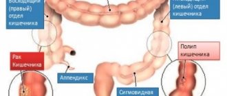
It is also worth considering that one of the most common causes of inflammation of the maxillary sinuses is poor oral hygiene and poor dental care, which includes delays in seeking dental care and refusal of proposed treatment.
People with tooth decay or other dental diseases have an increased risk of developing sinusitis and sinusitis (a disease in which inflammation affects the frontal sinuses).
One of the first signs indicating the onset of the inflammatory process is toothache, radiating to the temple and gradually spreading to the forehead.
Treatment of sinusitis (case study) –
The following example clearly shows a typical medical error, as a result of which patients, even if they consult a doctor in a timely manner, suffer from sinusitis for years. As you will see below: after eliminating errors in diagnosis, the treatment of sinusitis in this patient was carried out in full (using endoscopic methods).
A 28-year-old female patient came to the medical center with symptoms of unilateral acute purulent sinusitis. Computed tomography revealed that the sinus was half filled with pus (Fig. 15-16). The treatment that was prescribed was punctures and rinsing of the sinuses with antiseptics and massive antibiotic therapy. As a result, for the next 3 years the patient regularly visited this clinic with similar symptoms and was given similar therapy.
Unilateral acute purulent sinusitis on CT (different projections):
During this time, the attending physician never referred the patient to a dentist to determine how well the root canals in teeth 5-6-7 on this side were filled. Moreover, during this time the patient had CT images taken outside the exacerbation period. When the patient comes to me for a consultation, an image taken outside the exacerbation period reveals a round formation up to 1 cm in size in the area of the bottom of the maxillary sinus, which is not typical for the topography of the sinus (Fig. 17). My preliminary diagnosis is a maxillary sinus cyst on the right (it’s on the left in the CT image).
Own cysts of the sinus mucosa are extremely rare, and practically the only reason for their formation is inflammation in the area of the apex of the roots of one of the 5-6-7 upper teeth. In addition, the CT scan clearly showed that the apexes of the roots were located directly under the mucous membrane of the sinus (i.e., there was no layer of bone between the apexes of the roots and the bottom of the sinus). As a result, I refer the patient to “diagnostic endonasal maxillary sinusoscopy,” which involves the use of a thin flexible probe with a video camera. As a result, we see a cyst filled with pus, which is located in the projection of 5 or 6 teeth (Fig. 18). We were unable to accurately determine the causative tooth either from a CT scan or from a video (plus each of these teeth had problems with the quality of root canal filling).
As a result, the patient was initially referred for an endoscopic maxillary sinusotomy using a shaver (through the nose), a technique used to curettage and remove polyps and cysts in the sinus. During the operation, the patient also had the anastomosis between the maxillary sinus and nasal passages widened. 1.5 months after the operation and repeated diagnostic maxillary sinusoscopy, the patient was sent for retreatment of 5-6-7 teeth (including sanitation of the root canals using preparations based on calcium hydroxide). As a result, the patient is happy and satisfied.
Important: as an oral and maxillofacial surgeon, I sincerely recommend that (if the ENT doctor himself does not consider it necessary to refer you for a CT scan of your teeth, as well as for a consultation with a dentist), you independently consult a dentist-therapist for a consultation. So that a thorough examination of each root canal in the area of 5-6-7-8 teeth is carried out. Unfortunately, as we see in a specific example, ENT doctors very often make mistakes in diagnosing the odontogenic nature of the origin of sinusitis.
Folk remedies
Traditional medicine offers many options for treating sinusitis:
- Kalanchoe. It is necessary to wash the Kalanchoe leaves and chop them. You can use it in the same way as an onion lotion, or bury it, Kalanchoe will provoke sneezing, during which the nose and sinuses are cleared.
- Rosehip and sea buckthorn oils are instilled into the nose 6-8 times a day. This helps soften the nasal mucosa and improve the drainage of altered tissues.
- Beet. Beetroot juice has an antibacterial effect; it is enough to bury it in the nose several times a day.
- Aloe. Apply 2 drops of juice from large aloe leaves into each nostril. Aloe is known for its bactericidal properties.
- Rinse with salt water. Take half a liter of boiled water and a teaspoon of sea salt, mix thoroughly so that the solution does not damage the mucous membrane. Take any convenient vessel (bottle or teapot) and pour it into one nostril so that the water comes out freely from the other. You need to bend over and turn your head to the side. Washing should be done up to 4 times a day.
- An effective remedy for headaches that often accompany inflammation of the maxillary sinuses is cyclamen root juice. 2 drops of juice are instilled into both nostrils of the patient, while the patient is in a lying position. Five minutes later, the patient has a reaction in the form of sneezing, coughing and fever. After this procedure, pus is discharged from the patient’s nose for a whole day. Headaches are relieved and sleep patterns are normalized. There is also a pharmaceutical preparation based on the juice of cyclamen root - Sinuforte.
Decoctions of medicinal herbs are suitable for rinsing: chamomile, string, yarrow, coltsfoot; as well as an ordinary weak solution of table salt.
- Before rinsing, the nasal cavity is carefully cleaned of dried crusts, softening them with sea buckthorn oil.
- Then draw the solution into the nostril, tilt your head back, hold in this position for a while, and blow the solution out of your nose.
You can pour the solution by sucking it through one nostril and blowing your nose through the other.
Causes
Chronic sinusitis in the vast majority of cases is a consequence of acute inflammation of the maxillary sinuses or (much less often) other organs. In this case, pathogenic microorganisms can enter the sinus cavity in several ways:
- hematogenous - entry of pathogenic agents through the systemic bloodstream from the main focus;
- rhinogenic – spread of the inflammatory process from the middle nasal passage through the communicating opening into the sinus cavity;
- odontogenic – infection of the sinus due to inflammatory diseases of the dental system, dental procedures.
In addition, chronic sinusitis can transform from acute traumatic (resulting from trauma to the facial skull), and have an allergic or vasomotor nature.
Chronication of acute sinusitis is promoted by:
- incompetent treatment of acute sinusitis with persistence of residual effects after completion of the main treatment cycle;
- anatomical features [congenital narrowness of the nasal passages or the outlet of the maxillary sinus, bending (congenital or post-traumatic) of the nasal septum, proliferation of tissues that form the nasal turbinates], impeding the outflow of mucus during acute inflammation and provoking its stagnation;
- polyps, cystic formations in the nasal cavity;
- chronic inflammatory diseases of the ENT organs;
- the presence of an unsanitized chronic inflammation in the oral cavity;
- unfavorable environmental or microclimatic conditions in the area of residence (gas pollution, increased dust levels, dampness, etc.).
Chronic sinusitis is characterized by cyclical exacerbations, provoked by a wide range of external and internal factors:
- spring-autumn period (wet, cold weather);
- state of immunosuppression;
- general hypothermia;
- exacerbation of other chronic diseases, leading to a decrease in the body’s reactivity;
- acute stressors;
- physical stress; and etc.
Prevention
- Try not to contact people who have a cold. Don't forget to wash your hands.
- Allergy sufferers need to monitor their condition. Get advice from your doctor on how to avoid provoking the development of an allergic reaction and control symptoms.
- It is recommended to avoid cigarette smoke. Try not to come into contact with dirty air. These provocateurs irritate the upper and lower respiratory tract.
- Doctors advise purchasing special air humidifiers.