Indications for appendectomy
- acute or chronic appendicitis;
- condition after appendiceal infiltration;
- the presence of neoplasms of the appendix.
Emergency surgery is usually performed no later than 1 hour after an accurate diagnosis is made.
If we are talking about a previous appendiceal infiltrate or a chronic course of the disease, surgical intervention is performed as planned (within 2 months to six months). The operation may be delayed for some time in patients who are in a state of intoxication, preschool children, as well as in elderly patients. In acute appendicitis, there are no contraindications to appendectomy. The only condition when surgery cannot be performed is late agony.
If we are talking about a planned operation, the patient should be carefully examined. Direct contraindications to radical surgery may include acute and chronic pathologies of the heart, kidneys, lungs, and liver.
Contraindications
Traditional appendectomy has virtually no contraindications, but laparoscopic appendectomy may not be used in all cases. To perform an appendectomy safely, the doctor needs to assess the patient's condition. Deviations are possible in the following cases:
- More than 24 hours have passed since the onset of the disease. In such cases, abscesses and ruptures appear, and antibiotics may be needed for appendicitis.
- The presence of inflammatory processes in the digestive organs.
- Another contraindication is the presence of disorders in other organs (for example, the development of cancer). Why is this situation so dangerous? It can negatively affect the patient's health. This applies to diseases such as heart failure, destructive processes in the lungs and bronchi, myocardial infarction, etc.
As a rule, the appendix is operated on urgently and the operation is not preceded by preliminary preparation.
Preparing for surgery
An appendectomy can be performed as an emergency or planned surgical procedure. It all depends on what stage the inflammatory process is at, where the appendix is localized, and what size the abscess is, if any.
Abdominal surgery is started only when an accurate diagnosis is established. Emergency surgery is performed in the presence of life-threatening symptoms (peritonitis and increasing manifestations of sepsis).
If the patient asked for help himself, not urgently, it is possible to observe the patient and more carefully prepare for the upcoming appendectomy. It is advisable that the patient undergoes the full range of necessary diagnostic tests; this will make it possible to minimize the risk of most complications and select the optimal option for pain relief.
Standard preparation protocol
On the eve of an appendectomy, it is necessary to perform a number of mandatory preparatory manipulations and procedures:
- Examine the cardiovascular system (using an ECG);
- Choose the most appropriate anesthesia option;
- Prepare the abdominal area that will undergo surgery (shaving the hair on the surgical site);
- Conduct a series of laboratory tests (general blood and urine analysis, coagulogram, HIV test, syphilis, hepatitis);
- Conduct instrumental studies (ultrasound of the appendix, abdominal organs).
Symptoms and diagnosis
Typical symptoms of acute appendicitis:
- abdominal pain;
- nausea and vomiting;
- fever;
- gas formation;
- urinary disorders;
- rapid expiration of general terms and conditions.
The pain may initially spread throughout the abdomen, and then it is usually localized just above the right groin area (right iliac fossa); pain may vary depending on the location of the appendix, radiating to the hip and lumbar region.
In women, gynecological pathologies should be excluded, which can manifest themselves with clinical pictures and very similar symptoms.
When palpating the abdomen at the level of the appendix, pain occurs, accompanied by a more or less clear contraction of the muscle wall (protective contracture).
Treatment methods
The traditional scenario for the operation to remove the appendix is carried out by creating a small incision not exceeding 12 centimeters. The entire procedure can be divided into several stages:
- Introducing the patient into a state of anesthesia. Today, removal of the appendix is most often performed under general anesthesia. If there are contraindications to general anesthesia, anesthesia is performed using the tight infiltration method or through conduction blockade;
- After this, the surgeon dissects the abdominal wall layer by layer, this allows you to avoid postoperative complications associated with damage to nerve endings, as well as respond in a timely manner to the sudden occurrence of bleeding;
- The muscles, like the edges of the surgical wound, are separated by blunt surgical instruments;
- After opening the internal space of the abdominal cavity, the doctor carefully examines the abdominal wall itself, assesses the condition of neighboring organs and begins to remove the intestinal loops, behind which the appendix itself is located;
- Next, the surgeon removes the appendix and sutures the surgical wound. First, the inflamed process is isolated from other tissues using a clamp and ligature;
- The surgeon places a purse-string suture on the stump (the edges of the sutures are inside the stump);
- After completing all surgical procedures inside the abdominal cavity, the surgeon forms external sutures. The peritoneal walls are usually held together using self-absorbable suture material. The surgeon places from 8 to 12 stitches, using synthetic or silk threads;
- The external postoperative suture must be removed 1 - 2 weeks after surgery.
Prevention
Appendicitis is not a highly preventable condition, although several studies have shown how lifestyle and certain types of food may protect against appendicular inflammation; in particular, it is recommended to follow a healthy and balanced diet rich in fiber and easily digestible foods, in addition to drinking enough fluids and moderate physical activity, which helps regulate intestinal motility.
On the contrary, excessive amounts of fatty foods, poorly digestible and rich in waste, especially if in conditions of constipation and intestinal irregularity, can contribute to inflammation of the appendix.
If appendicitis is suspected or confirmed, the following should be avoided:
- fried and fatty foods (sweets, sausages, fatty and fermented milk cheeses);
- foods that cause intestinal swelling due to gas production (broccoli, beans);
- alcoholic drinks, carbonated drinks, coffee and tea;
- spices, pepper and spicy seasonings.
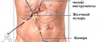
Laparoscopy
Laparoscopic surgery to remove the appendix is considered one of the most popular methods of removing the appendix. Surgical intervention is performed through micro-incisions. Using high-tech endoscopic equipment, the surgeon gives himself access to the abdominal cavity to the inflamed appendix.
At the initial stage of the operation, a gas mixture is introduced into the abdominal cavity. A miniature camera is inserted into one of the punctures, which will transmit the image to the monitor. The surgeon will be able to see everything that happens in the closed abdominal cavity. Then all the same manipulations occur as with the classic method of surgical removal of the appendix.
Minimally invasive techniques for removing the appendix
Despite the fact that classic appedectomy and laparoscopic removal of the appendix of the cecum are used quite often, the popularity of minimally invasive techniques is increasing every day. We are talking about the following forms of surgical intervention:
- transgastric appendectomy. It is performed without external incisions. To gain access to the tissues of the cecum and enter the abdominal cavity, a system of flexible instruments is used. The devices are inserted through the digestive tract, passing through the desired area of the intestine;
- transvaginal appendectomy. Access to the source of inflammation is carried out along an ascending route through a micro-incision in the vaginal wall. When choosing this method for removing the appendix, the location of the lesion plays a significant role.
Operations of this type make it possible to prevent complications associated with direct tissue trauma. They cannot be carried out if peritonitis is suspected, in the presence of multiple large foci with inflammation, or when the patient has manifestations of sepsis.
Acute appendicitis
In surgery, there is no disease more famous than acute appendicitis, but at the same time, in most cases, its recognition is difficult. This is due, on the one hand, to the fact that appendicitis, especially in the early stages of the disease, often does not have characteristic clinical manifestations, and on the other hand, to the fact that the doctor observing the patient has limited time to recognize the disease and choose treatment tactics. The latter circumstance excludes the use of any complex instrumental (except ultrasound) and laboratory techniques, therefore the diagnosis of acute appendicitis is based almost exclusively on data from questioning and physical examination.
History and general examination play an extremely important role in the diagnosis of acute appendicitis. The onset of pain in the epigastric region or throughout the abdomen with a gradual shift to the right iliac region (Kocher-Volkovich symptom) is characteristic of acute appendicitis and is rarely found in other diseases. Equally characteristic of appendicitis is scanty one- or two-time vomiting, which usually occurs in patients at home before admission to the surgical hospital.
An objective examination should first of all assess the general condition. It suffers little in case of catarrhal and phlegmonous appendicitis, but in its gangrenous form, despite the optimistic mood of the patient, it is possible to identify pallor, immobility, dry coated tongue, tachycardia and other signs of a systemic inflammatory reaction. The general condition suffers even more with perforated appendicitis. The patient is usually pale, indifferent, and sometimes groans in pain. Facial features are sharpened, knees are often brought to the stomach, pulse is significantly increased, blood pressure is lowered.
Examination of the abdomen in the catarrhal form of acute appendicitis does not reveal any features, but in the phlegmonous form it is possible to find some lag in the right iliac region during breathing. In the gangrenous form, this lag becomes easily noticeable, and in perforated appendicitis, the entire right half of the abdomen does not participate in breathing.
Palpation of the abdomen should be carried out carefully, otherwise incorrect data may be obtained due to the active resistance of the patient. First, superficial palpation is performed, which begins from the left iliac region, then, gradually moving to the right, a zone of hyperesthesia and local tension of the abdominal muscles in the right iliac region are identified.
After this, methodical deep palpation is performed according to Obraztsov-Strazhesko, which also begins with the left lower abdomen, and then, through deep step-by-step palpation, tenderness is detected in the right iliac region. With empyema of the appendix in thin people, it is possible to palpate the thickened appendix in the form of an elongated, mobile, painful formation. It should be remembered that deep palpation in destructive forms of appendicitis is often impossible due to pronounced pain and muscle tension.
By palpation, the symptoms of Voskresensky, Shchetkin-Blumberg, Rovzing, Sitkovsky, Bartomier-Mikhelson are first determined, and then the Obraztsov symptom. We must not forget about palpation of the right lumbar region, which can be crucial for recognizing the retrocecal form of acute appendicitis. In this case, soreness, muscle tension and the Shchetkin-Blumberg symptom will be determined accordingly in this zone.
Vaginal and rectal examinations are strictly required. Their value lies in the fact that they provide direct palpation of the lowest floor of the abdominal cavity. If the appendix is located in the pelvis or if there is an accumulation of purulent effusion here, these studies will reveal significant pain in the area of the posterior vaginal vault or the anterior wall of the rectum, as well as the characteristic fluid discharge. In addition, vaginal examination can play a decisive role in the differential diagnosis between acute appendicitis and inflammation of the uterine appendages.
The minimum tests required to establish a diagnosis of acute appendicitis include measuring body temperature and determining the leukocyte reaction. The temperature, as a rule, is elevated in any form of acute appendicitis, but it rarely exceeds 38 ° C. Only with a developing appendiceal abscess or diffuse peritonitis can the body temperature reach or exceed 39 ° C.
Leukocytosis is characteristic of all stages of acute appendicitis. We described the dynamics of its changes when covering the clinical forms of the disease. It is only necessary to add that in the diagnosis of acute appendicitis, leukocytosis has no independent significance, since it is also observed in other inflammatory diseases, and with the development of gangrene of the appendix, it may not be present at all. Much more important is the assessment of the white blood formula, in particular the presence or absence of a neutrophil shift: an increase in the content of young forms (band neutrophils, myelocytes and promyelocytes) to the detriment of the content of mature (segmented) neutrophils. With the rapid development of the destructive-inflammatory process, the total number of leukocytes can even be reduced (and sometimes significantly!), while young forms of neutrophils predominate in the white blood formula. This so-called leukocytosis of consumption reflects the extreme degree of strain on the hematopoietic system, when it is not able to fully provide the body’s needs during inflammation with the proper number of neutrophils. Therefore, in itself, a general decrease in the number of leukocytes in the peripheral blood against the background of a pronounced clinical picture of acute appendicitis cannot serve as a sign of the subsidence of the inflammatory process. Only an assessment of changes in the leukocyte formula can create a true idea of the positive or negative dynamics during an attack of acute appendicitis.
The diagnosis of acute appendicitis with a typical localization of the appendix is most difficult during the first hours of the disease, i.e. in the stage of “catarrhal” inflammation, since during this period the symptoms are nonspecific: the general condition of the patient suffers little, there are no symptoms of peritoneal irritation, etc. That is why With the catarrhal form of acute appendicitis, the largest number of diagnostic errors occurs. At best, this leads to unnecessary removal of the appendix. However, careful assessment of the history and physical examination, as well as the use of laparoscopy, greatly reduce the incidence of misdiagnosis and thus eliminate unnecessary appendectomies.
In the initial stage of the disease, when there are no signs of a destructive process and the diagnosis of acute appendicitis is in doubt, dynamic monitoring of the patient until clear manifestations of the disease appears. During this period, special studies can be carried out to facilitate the differential diagnosis: chromocystoscopy, excretory urography, laparoscopy, ultrasound scanning of the abdominal cavity.
Differential diagnosis
Acute appendicitis must be differentiated from almost all acute diseases of the abdominal cavity and retroperitoneal space. This, on the one hand, is facilitated by the extreme variability in the location of the appendix, and on the other hand, by the frequent absence of specific symptoms of the disease.
In the initial stage, when the pain does not yet have a clear localization in the right iliac region, but is observed mainly in the epigastric or mesogastric region, it is most often necessary to differentiate acute appendicitis from acute gastroenteritis, acute pancreatitis, and much less often from perforation of a stomach and duodenal ulcer.
Acute gastroenteritis, unlike acute appendicitis, begins with fairly severe cramping pain in the upper and middle parts of the abdomen. When questioned
The patient is almost always able to identify the provoking factor in the form of a change in diet, for example, taking a large amount of fatty and spicy foods, alcohol, etc. Almost simultaneously with cramping pain, repeated vomiting occurs, first of the food eaten, and then of bile. With significant damage to the gastric mucosa, vomiting mixed with fresh blood may occur. After a few hours, frequent loose stools often appear against the background of cramping pain.
During an objective examination of the abdomen, attention is drawn to the absence of localized pain, symptoms of peritoneal irritation and symptoms characteristic of acute appendicitis: Rovzing, Sitkovsky, Obraztsov, etc. During auscultation of the abdomen, increased peristalsis is heard. Digital rectal examination reveals the presence of liquid stool mixed with mucus, the absence of overhang and pain in the anterior wall of the rectum. Body temperature is usually normal or subfebrile. Leukocytosis increases moderately, band shift is absent or slightly expressed.
Acute pancreatitis, unlike acute appendicitis, begins with sharp pain, often of a girdling nature, in the upper abdomen. The pain often radiates to the back and is accompanied by repeated vomiting of bile, which does not bring relief.
In the initial stage of acute pancreatitis, patients behave restlessly, then, as intoxication increases, they become lethargic and adynamic; with a rapidly progressing disease, collapse may occur. The skin is pale, sometimes with some acrocyanosis, the pulse is significantly increased, while the temperature, at least during the first hours of the disease, remains normal.
During an objective examination of the abdomen, attention is drawn to the discrepancy between the severity of the general condition and the relatively unexpressed pain in the epigastric region. In the right iliac region there is most often no pain at all. Only in the late stages of acute pancreatitis, as the effusion spreads from the omental bursa and the right hypochondrium towards the right lateral canal and the iliac region, symptoms simulating acute appendicitis may appear. Meanwhile, in this case, the history of the disease, the presence of maximum pain in the epigastric region and symptoms characteristic of acute pancreatitis (absence of pulsation of the abdominal aorta in the epigastrium, the presence of painful resistance of the abdominal wall slightly above the navel and pain in the left costovertebral angle) will help to establish true diagnosis.
In difficult cases of differential diagnosis, laboratory assessment of the content of amylase (diastase) in the blood and urine is of great help. In particular, if the amylase content in the urine exceeds 128 units. (according to Wolgemut), then if there is doubt about the diagnosis, this fact indicates rather in favor of acute pancreatitis.
A perforated ulcer of the stomach or duodenum has such a characteristic clinical picture that it is almost difficult to mistake this complication of a peptic ulcer for acute appendicitis. The presence of the classic triad (gastric history, dagger pain in the epigastric region, widespread muscle tension), as a rule, allows you to immediately establish an accurate diagnosis. In addition, when an ulcer is perforated, vomiting is very rare and the disappearance of hepatic dullness is often detected - a symptom pathognomonic for perforation of a hollow organ.
Diagnostic doubts arise only in cases of covered perforation of the ulcer, when the stomach contents that have entered the abdominal cavity and the resulting effusion gradually descend into the right iliac fossa, where they are retained. Accordingly, the pain shifts: it subsides in the epigastric region after covering the perforation and, conversely, arises in the right iliac region as gastric contents penetrate into it. Such a false Kocher-Volkovich symptom can contribute to an incorrect conclusion about the presence of acute appendicitis and lead to an error in surgical tactics. The error is all the more possible because in the described situation, muscle tension and other symptoms of peritoneal irritation are clearly visible in the right iliac region: Shchetkin-Blumberg, Voskresensky, etc. Based on this, in this case, the assessment of the immediate and long-term history of the disease becomes extremely important. Long-term gastric discomfort or direct indications of a previous peptic ulcer, the onset of an acute disease not with dull, but with very sharp pain in the epigastric region, and the absence of vomiting do not indicate acute appendicitis. Doubts can be completely resolved by percussion or x-ray detection of free gas in the abdominal cavity.
Unfortunately, in a number of cases of covered perforation of an ulcer, simulating acute appendicitis, it is not possible to avoid a diagnostic error and a true diagnosis becomes possible only during surgery.
With an unusual localization of the appendix (under the liver, near the urinary tract, in the small pelvis), there is a need for a differential diagnosis between acute cholecystitis, urological and gynecological diseases.
Acute cholecystitis, unlike acute appendicitis, most often begins not with dull, but with very sharp pain in the right hypochondrium with typical irradiation to the right shoulder and scapula. This initial stage of acute cholecystitis, known as biliary (hepatic) colic, is often accompanied by repeated vomiting of food and bile.
When questioning the patient, as a rule, it turns out that such attacks of pain occurred more than once and their appearance was associated with a change in the usual diet: taking large amounts of fatty foods, smoked foods, and alcohol. Sometimes a history can establish the presence of transient jaundice that occurs shortly after an attack of pain. These anamnestic indications indicate acute cholecystitis.
When examining the abdomen, it should be taken into account that in the case of a high position of the appendix, maximum pain and muscle tension are localized in the lateral parts of the right hypochondrium, while with cholecystitis these signs are revealed more medially. In acute cholecystitis, it is often possible to palpate an enlarged and sharply painful gallbladder.
Body temperature in acute cholecystitis is significantly higher than in acute appendicitis in all stages of the disease, although in general the destructive process in acute cholecystitis develops more slowly than in appendicitis. As a rule, there is no significant difference in the dynamics of leukocytosis in both cases.
Valuable information can be obtained from ultrasound examination. It allows you to clearly visualize the gallbladder and detect signs typical of its inflammation (increase in the volume of the bladder, the thickness of its walls, layering of the walls, etc.). At the same time, hopes for ultrasound verification of the diagnosis of acute appendicitis are unjustifiably exaggerated, although this sometimes fails.
Right-sided renal colic, as a rule, begins not with dull, but with extremely sharp pain in the right lumbar or right iliac region. Often, against the background of pain, vomiting occurs, which is of a reflex nature. Pain in typical cases radiates to the right thigh, perineum, genitals and is accompanied by dysuric disorders in the form of frequent and painful urination. It should be noted that dysuric phenomena can also be observed in acute appendicitis if the inflamed appendix is in close proximity to the right kidney, ureter or bladder, but in this case they are less pronounced. In the differential diagnosis, an extremely important role is played by the anamnesis: as is known, with appendicitis there is never very severe paroxysmal pain with the irradiation described above. In addition, despite strong subjective pain, during a physical examination of a patient with renal colic, it is not possible to detect either intense pain in the abdomen or symptoms of peritoneal irritation.
Doubts can be resolved after a laboratory urine test and, if possible, urgent emergency urography or chromocystoscopy. Sometimes even macroscopically it is possible to note the intense red color of urine (macrohematuria), not to mention the fact that microscopy of urinary sediment almost always reveals an excess content of fresh red blood cells (microhematuria). Detection of a violation of the passage of urine in one of the ureters, established by urography or chromocystoscopy, in the presence of the pain syndrome described above, serves as a pathognomonic sign of renal colic. Sometimes routine radiography of the urinary tract helps to resolve diagnostic doubts, in which it is possible to notice the shadow of a radiopaque stone. An ultrasound examination brings some clarity to the diagnosis, revealing in a number of patients stones in the projection of the right ureter and an increase in the size of the right kidney.
Right-sided pyelitis (pyelonephritis) in relation to the differential diagnosis with acute appendicitis is a more difficult task than renal colic. The disease, as a rule, begins subacutely with dull arching pain in the lumboiliac or mesogastric region. Vomiting and dysuria are often absent at the onset of the disease. Only after 1-2 days does the body temperature rise sharply (up to 39°C and above). Of course, in this case, the medical history plays an important role, since pyelitis is most often a consequence of impaired urination as a result of urolithiasis, pregnancy, prostate adenoma, etc.
Even in the presence of obvious signs of purulent intoxication, it will not be possible to detect sharp pain on palpation of the abdomen and symptoms of peritoneal irritation. At the same time, it should be noted that with pyelitis, pain in the mesogastric region, iliac region and a positive psoas-symptom of Obraztsov is often determined.
As with renal colic, urine testing plays an important role in the differential diagnosis of pyelitis and acute appendicitis, which can reveal pyuria. Survey and contrast urography for pyelitis are less important than for renal colic, although they often reveal unilateral or bilateral pyelectasis in the patient, which can also be determined by ultrasound.
Features of the differential diagnosis of acute appendicitis and gynecological pathology are presented in Chapter XVIII.
Clinical manifestations of other acute but rare diseases of the abdominal organs: terminal ileitis (Crohn's disease) in its initial stage, Meckel's diverticulitis , etc. do not have any characteristic differences from the signs of acute appendicitis, therefore the correct diagnosis is most often established only during operations.
A great help in difficult cases of differential diagnosis in acute pathology of the abdominal organs is laparoscopy, which is becoming increasingly widespread in surgical hospitals. Laparoscopic examination should be carried out in the operating room in such a way that, if necessary, emergency surgery, including laparoscopic appendectomy, can be started immediately. From the same point of view, it is advisable to perform laparoscopy under general anesthesia.
The differential diagnosis between acute appendicitis and right-sided pyelitis in the second half of pregnancy presents certain difficulties, since the diseases have many similar features: mild pain, high localization of pain, and the presence of vomiting. As for the dysuric phenomena inherent in pyelitis, they can also be observed in acute appendicitis due to the close proximity of the appendix and the right kidney in late pregnancy.
An objective examination of the abdomen indicates the presence of highly localized tenderness, and since the ileocecal angle is located behind the sharply enlarged uterus, symptoms of peritoneal irritation may not be detected for a long time.
Only the onset of the disease is significantly different: it is known that appendicitis always begins with pain, followed by an increase in body temperature and vomiting, while with pyelitis the disease most often begins with severe chills, vomiting, an increase in temperature, and only after this does pain occur. With a careful objective examination, it is possible to notice that the maximum pain in the case of pyelitis in pregnant women is closer to the lumbar region, while in case of appendicitis it is detected in the area of the lateral and anterior wall of the abdomen. Palpation of the abdomen with the patient on the left side can help in correct orientation; in this case, due to some displacement of the uterus to the left, it is possible to palpate the area of the vermiform appendix and the right kidney in more detail. It is also important to examine urine (necessarily taken with a catheter!), which can detect the presence of pyuria as a sign of purulent pyelonephritis, as well as an ultrasound examination that reveals changes in the pyelocaliceal system of the right kidney. However, if doubts remain, it is better for the patient to undergo surgery rather than undergo conservative treatment with the risk of developing appendiceal peritonitis. In pregnant women, surgical tactics should be more active than in other categories of patients.
With regard to pain relief in pregnant women, the rule remains the same as outside pregnancy: for any form of acute appendicitis, general anesthesia should be preferred.
As a surgical approach for an undoubted diagnosis in the first half of pregnancy, the Volkovich-Dyakonov incision is used. In the second half of pregnancy, this access may be inadequate, so it is modified according to the principle: the longer the pregnancy, the higher the incision. Thus, in the last weeks of pregnancy, the incision is made above the ilium due to a significant upward displacement of the cecum and appendix. It is advisable to expand the Volkovich-Dyakonov incision by incising the edge of the sheath of the right rectus abdominis muscle.
Surgical tactics for any form of appendicitis in pregnant women do not differ from the generally accepted principles of its treatment. In other words, the features of the surgical technique and methods of drainage of the abdominal cavity, adopted for various forms of acute appendicitis, fully retain their significance here. It is only necessary to exercise maximum caution when manipulating near an enlarged uterus, since trauma to it can be a direct cause of miscarriage or premature birth. For the same reasons, abdominal tamponade is carried out according to the strictest indications: 1) if it is impossible to achieve reliable hemostasis in the abdominal cavity; 2) when opening a circumscribed periappendiceal abscess.
In the postoperative period, in addition to conventional therapy, it is necessary to prescribe treatment aimed at preventing premature termination of pregnancy. Strict bed rest is prescribed, administration of a 25% solution of magnesium sulfate 5-10 ml 2 times a day intramuscularly, administration of tocopherol acetate at a dose of 100-150 mg per day as an injection of a 10% oil solution 1 ml 1 time. In the absence of laboratory monitoring of hormonal levels, the prescription of hormonal drugs (progesterone, etc.) should be avoided, since in some cases an overdose can have the opposite effect. The administration of proserin and hypertonic sodium chloride solution as agents that promote uterine contraction is strictly contraindicated. For the same reason, hypertensive enemas should not be used.
In pregnant women, the most difficult task is the treatment of diffuse peritonitis. Mortality from this complication remains very high and, according to various authors, is 23-55% for the mother and 40-92% for the fetus, with the highest mortality observed in late pregnancy. Unfavorable results of treatment of diffuse purulent peritonitis in pregnant women gave rise to extreme radicalism in surgical tactics. It was considered necessary to perform the following volume of surgical intervention: immediately after opening the abdominal cavity, perform a cesarean section, then supravaginal amputation of the uterus, then appendectomy, toilet and drainage of the abdominal cavity. Currently, thanks to the availability of powerful antibacterial agents, in most such cases it is possible not to resort to a cesarean section, much less subsequent amputation of the uterus. It must be emphasized that the question of the volume and nature of intervention for destructive appendicitis against the background of long periods of pregnancy should be resolved together with an obstetrician-gynecologist, with his direct participation in surgical intervention. Briefly, the principle of modern surgical tactics can be formulated as follows: maximum activity in relation to peritonitis, maximum conservatism in relation to pregnancy.
In modern conditions, with diffuse appendicular peritonitis in pregnant women, a median laparotomy, evacuation of pus, appendectomy, abdominal toilet, and drainage are installed under general anesthesia. The surgical wound is sutured tightly. In case of full-term or almost full-term pregnancy (36-40 weeks), due to the inevitability of childbirth against the background of peritonitis, the operation begins with a cesarean section, then after suturing the uterus and peritonization of the suture, an appendectomy and all further manipulations associated with the treatment of peritonitis are performed.
The urgent need for amputation of the uterus arises only when it is destructively damaged, which is occasionally observed in conditions of diffuse purulent peritonitis. It should also be taken into account that with diffuse purulent peritonitis, the contractility of the uterus is significantly reduced. In this regard, sometimes after a cesarean section there is a danger of atonic bleeding, the only way to combat which is immediate amputation of the uterus.
Acute appendicitis during childbirth deserves special attention In the literature, there are conflicting opinions about when in such cases it is necessary to perform an appendectomy: before or after delivery, whether to allow childbirth to proceed naturally, or to perform a cesarean section and appendectomy at the same time. In this regard, it should be noted that surgical tactics for appendicitis during childbirth depend both on the course of labor and on the clinical form of acute appendicitis. So, if childbirth proceeds normally with the clinical picture of catarrhal and phlegmonous appendicitis, then it is necessary to promote the fastest natural delivery and then perform an appendectomy. If, against the background of the normal course of labor, there is a clinical picture of gangrenous or perforated appendicitis, then it is necessary to temporarily stop the contractile activity of the uterus, perform an appendectomy and then again stimulate labor. In conditions of pathological childbirth, it is necessary to perform a simultaneous cesarean section and appendectomy for any clinical form of acute appendicitis.
Regardless of the timing of delivery, the patient must be transferred to the surgical department for appendectomy and subsequent postoperative management, where she must be observed by both a surgeon and a gynecologist.
Acute appendicitis in children is much less common than in adults. The vast majority of cases occur over the age of 5 years. The rarity of acute appendicitis before the age of 5 is explained by the fact that the appendix has a funnel-shaped shape, which facilitates good emptying of the appendix, and also by the fact that the lymphoid apparatus of the appendix is still poorly developed during this period of life.
Acute appendicitis in children occurs more violently than in adults. This is due to the child’s body’s insufficient resistance to infection, weak plastic properties of the child’s peritoneum, and insufficient development of the omentum, which does not reach the appendix and thus does not participate in the creation of a delimiting barrier.
The pain that arises in the abdomen is often cramping in nature and does not have the clear dynamics that are characteristic of acute appendicitis in adults. It should be noted that children under 10 years of age, as a rule, cannot accurately localize pain, which makes it difficult to recognize the disease. Vomiting in children is most often repeated, stools do not tend to be delayed, and in young children they even become more frequent. The posture of a sick child is characteristic. He lies on his right side or on his back, bringing his legs to his stomach and placing his hand on the right iliac region, protecting it from examination by a doctor. With careful palpation, it is often possible to identify hyperesthesia, muscle tension and the area of greatest pain. Even in the first hours of the disease, symptoms of Shchetkin-Blumberg, Voskresensky, Bartomier-Mikhelson are pronounced.
The temperature from the very beginning of the disease is significantly higher than in adults, often reaching and exceeding 38°C. The number of leukocytes is also increased, but it rarely exceeds 20x109/l along with the existing neutrophil shift.
In the differential diagnosis of acute appendicitis in children, the following diseases deserve attention: pleuropneumonia, acute gastroenteritis, dysentery, hemorrhagic vasculitis (Henoch-Schönlein disease).
When differentiating pleuropneumonia , it must be taken into account that this disease is characterized not only by pain spreading towards the abdomen, but also by cough, sometimes with transient cyanosis of the lips, wings of the nose and shortness of breath. It should be recalled that in children the normal ratio of breathing to pulse is 1:4, and if
the ratio becomes 1:3 or 1:2, then this rather speaks in favor of acute pneumonia. With pleuropneumonia, you can also listen to rales and pleural friction sounds on the corresponding side of the chest.
When differentiating gastroenteritis , it must be taken into account that this disease begins, as a rule, not with abdominal pain, but with vomiting and the appearance of characteristic repeated watery stools; pain, unlike acute appendicitis, comes later. In addition, with gastroenteritis they have a pronounced cramp-like character, often followed by the urge to stool. The temperature in this disease is elevated, as in appendicitis, but the number of leukocytes is normal or even slightly reduced, and the neutrophil shift is not pronounced.
The need to differentiate acute appendicitis from dysentery occurs most often in the younger age group. Here, first of all, the anamnesis plays a role, in particular, indications that a similar disease appeared in several children at once, especially in children's groups. The pain in dysentery is clearly cramping in nature and is localized mainly in the left half of the abdomen; repeated loose stools are often accompanied by blood. Maximum palpation pain is determined in the lower abdomen on the left; symptoms of peritoneal irritation, with rare exceptions, are not detected. Body temperature during dysentery is often high (38.0-39.0 ° C), the number of leukocytes can be increased without a significant neutrophil shift.
To exclude hemorrhagic vasculitis , take into account that abdominal pain in this disease is caused by multiple small subserous hemorrhages and does not have a clear localization. In addition, a careful examination of the skin allows us to identify the presence or residual effects of hemorrhagic exanthema in symmetrical areas of the torso, limbs, and buttock areas. You should also pay attention to the mucous membrane of the cheeks and sublingual space, where it is possible to detect the presence of small submucosal hemorrhages even before the appearance of a rash on the skin. The abdominal wall is not tense during the examination, but the Shchetkin-Blumberg symptom is most often pronounced, the abdomen is distended and uniformly painful. A rectal examination may reveal bloody intestinal contents. Body temperature sometimes reaches 38°C and higher, the number of leukocytes is also most often increased without a significant neutrophil shift.
In case of significant difficulties in the differential diagnosis, if there are no symptoms of peritoneal irritation, dynamic observation of the child for 6-12 hours is acceptable.
At the same time, it should be remembered that appendicitis in children occurs much more violently than in adults, and often within the first day of the disease destruction of the appendix develops. Based on this, surgical tactics in children should generally be more active than in adults.
All this fully applies to appendicular infiltrate, which in children often forms already on the 2nd day of the disease. Since in children the appendix is relatively long, and the omentum, on the contrary, is short and the peritoneum does not have sufficient plastic properties, the resulting infiltrate is “fragile” and cannot serve as a reliable obstacle to the spread of infection throughout the abdominal cavity. In this regard, the operation is indicated even with a forming (palpable) appendiceal infiltrate, especially since isolating the appendix from loosely fused organs is not particularly difficult.
Appendectomy in children is always performed under general anesthesia. The Volkovich-Dyakonov incision is used as an operative access, with the exception of cases of diffuse purulent peritonitis, when a lower-median laparotomy is indicated.
In most cases, appendectomy in children is not technically difficult due to the absence of adhesions.
Early recovery period
After the operation is completed, the patient is observed for several days by the operating surgeon. Sutures are removed 7-10 days after surgery.
Early postoperative rehabilitation includes the following points:
- Detoxification of the body (in this case, activities are carried out both on the first day after surgery and in the following days);
- Following a strict diet;
- Restoring the functional potential of the intestines and bladder.
In the postoperative period, the patient may be prescribed medications (antibiotics, analgesics). Particular attention is paid to the prevention of constipation (for this purpose, a special diet and laxatives are prescribed).
Recovery
Analgesics
During the recovery period after surgery, the patient is prescribed painkillers.
The patient's postoperative condition is often accompanied by pain, especially at the suture. To make the patient feel less pain, medications are prescribed that will help numb the wound. Such analgesic medications are tablets and intramuscular injections. During the inpatient period, injections are more often used, and treatment can be continued at home with the help of tablet analgesics.
Motor mode
How long it will take to heal at the incision site depends on the type of appendectomy performed and the individual characteristics of the patient’s body. If you have anemia and diabetes, you will have to limit your physical activity; there is no need to run during this period. Until the healing process is complete, it is necessary to support the abdominal area during periods of laughter and coughing, moving carefully so that the abdominal muscles do not tense. At first it will be useful to stay in bed, and then gradually start walking and increasing the load. In addition, physiotherapy procedures are prescribed, such as: UHF therapy and laser treatment. These treatments will promote healing.
Diet therapy
During the rehabilitation period after appendectomy, special nutritional conditions are recommended. In the very first days, you need to limit your diet. Liquid low-fat products are suitable: yoghurts, cereals, jelly. The body will require more water. You should not eat foods that promote flatulence - peas, beans, kvass, cabbage, lentils, milk, etc. Food with excess fat and spices will also be harmful. Subsequently, fruits and vegetables, poultry and fish can be introduced into the diet. It is better to cook food by baking or steaming to make the task easier on the stomach. You can return to your usual diet in 2-3 weeks.