Cleft lip is a congenital malformation in which a newborn suffers from partial or complete underdevelopment of the upper lip. According to statistics, about 700 children with congenital facial clefts are born every day in the world. The pathology is formed in utero between 4 and 12 weeks of pregnancy.
The disease is characterized not only by local anatomical changes, but by accompanying systemic disorders. Children with a cleft lip cannot breathe, swallow, eat or speak normally.
General information
Cleft palate and lip (another name for this pathology is cheiloschisis , but colloquially it is called “cleft lip and cleft palate” ) is a congenital defect that forms during the fetal development of the baby, when, due to the influence of certain factors, the continuity of the upper lip and palate is disrupted and alveolar process.
Children who are born with this pathology have external facial deformation and significant functional impairment. It is very difficult for them to eat and learn to talk. Intellectually, such children develop normally.
However, the causes of the disease “cleft palate and cleft lip” may be associated not only with impaired intrauterine development. The acquired form of the disease can develop as a result of infectious processes, tumors, or physical damage. However, such cases are rare.
An effective treatment method is surgical correction. It is important, immediately after the birth of a baby with such a pathology, to provide him with observation by a highly qualified specialist and promptly carry out the correct treatment.
Treatment
Treatment of cleft lip is carried out through one or even several plastic surgeries, depending on the characteristics of each type of defect. Surgical correction of congenital cleft lip is performed for children who were born at term and do not have any contraindications (birth injuries, congenital defects of vital organs, acquired diseases, etc.). Surgical treatment of cleft lip should best be carried out at the age of 3-6 months. In the case of a more severe defect, it is permissible already in the first days or month of the baby’s life, provided there is sufficient weight gain, as well as the absence of anemia, pathology of the cardiovascular, digestive, nervous and endocrine systems.
There are three types of plastic surgery aimed at correcting this problem.
Cheiloplasty - this type of treatment is the simplest because it does not require correction of any other tissues. During the healing period of the sutures, a cotton swab is inserted into the nose in order to in no case allow tissue fusion. Usually 10 days after surgery, the sutures are removed.
Rhinocheiloplasty is a more severe type of surgical treatment, when in addition to an aesthetic result, it is necessary to achieve functional convenience. This operation is performed both on the affected lip itself and on the muscles of the facial region.
Rhinocheilognatoplasty is the most complex type of operation, which includes the previous two types, but also with correction of the respiratory canals.
Carrying out surgical treatment in the first two weeks of a child’s life has a beneficial effect on the further development of the lip and nose, and also significantly reduces the anxiety of parents. But it should be taken into account that at this age children have too small anatomical dimensions of the lip, some of their functions are imperfect, and the tendency to blood loss is significantly increased.
With the surgical removal of a cleft lip, restoration of its integrity and anatomical structure, correction of deformities of the nose, palate and other facial defects is achieved.
Correction of cleft lip should be completed by the age of three, when speech begins to develop. Subsequently, speech therapy treatment is carried out, which is aimed at eliminating speech defects, as well as cosmetic treatment in order to eliminate the postoperative scar.
Pathogenesis
The disease “cleft palate” develops as a result of a delay in the process of fusion of those bones that form the bony palate of the oral cavity - the maxillary processes with the vomer. Because of this, a cleft remains on the hard palate. Soft tissues also diverge, sometimes the process involves the lip. This syndrome is called “cleft lip” because in hares and rabbits the upper lip consists of two halves.
As a rule, pathology develops in the fetus during intrauterine development. Therefore, the disease can be detected even before birth by performing an ultrasound examination. The cleft appears in the embryo in the first months of its development, when the formation of the maxillofacial organs occurs. The bones in the area of the hard palate fuse in the fetus by the 11th week of development, and by the end of the third month the soft palate is formed. In general, the formation of the palate occurs at 2-3 months of intrauterine development. The lower lip is formed in the fetus at the end of the first month, when the mandibular processes fuse. The final formation of the upper lip occurs at the end of the second month, when the right and left maxillary processes fuse with the median nasal process. If during this period there is a negative influence of endogenous and exogenous, as well as genetically determined factors, this process goes wrong, and the baby develops congenital facial deformities - cleft lip and cleft palate.
The localization of such nonunions and their shape depends on where exactly the fusion did not occur. Most often, the upper lip and palate do not fuse; in more rare cases, the mandibular processes, maxillary and frontal processes, maxillary and mandibular germinal processes, medial nasal and frontal germinal processes do not fuse.
Where does cleft lip come from?
You will be able to better understand how facial defects are formed if you consider how a multicellular embryo is formed from 1 maternal and 1 paternal cell, and how it acquires a face.
So, after the fusion of the egg and the male cell, one cell is formed, which begins to divide into 2, then 4 and then cells, all of them identical. At the beginning of division, the cells become smaller and smaller, but after time the volume of the embryo begins to grow. Over time, as the cell mass grows, the following significant event occurs: the cells from which the body of a developing person is built become different. They form 3 layers:
- internal - endoderm, from the cells of which the intestines, liver, lungs, pancreas are subsequently formed;
- external - ectoderm, giving rise to skin, nails, hair, nervous system and sensory organs;
- middle - mesoderm, from which muscles, bones, blood vessels, heart, genitals and kidneys develop.
At 2 weeks of life, a depression forms between the developing brain and the area from which the heart will be formed - the primary mouth. It connects to a cavity within the endodermis called the primitive gut. This is how the digestive canal is formed.
At week 4, longitudinal impressions appear on one and the other side of the primary mouth - gill arches. There are 4 of them on each side, and they deepen so that structures similar to mounds are obtained. The anterior ends of the I and II gill arches bifurcate, forming several processes resembling petals. From the first gill arch, which gives rise to the entire face, there are 5 of them:
- 1 nasofrontal process;
- 2 maxillary;
- 2 mandibular.
Between the nasofrontal and maxillary processes there is a gap where the eye sockets will later be. In the space between the maxillary and mandibular processes, the mouth will form, and when they join in the lateral sections, the cheeks are formed. The anterior third of the auricle will also form from 1 gill arch.
The frontal and maxillary “petals” move towards each other first, then the cheeks, upper and lower jaws are formed: the skin, mucous membranes, salivary glands, and tooth enamel are formed from the ectoderm; from the mesoderm - bones and muscles of the face, internal parts of the teeth. If fusion of the processes does not occur, which can happen in any one or several places, the face changes. The severity of this change varies from a small cleft in the upper lip to a completely disfigured face.
Starting from the 4th to the end of the 8th week of intrauterine development, these processes must unite, but their complete fusion occurs until the 11th week inclusive. It turns out that in the period from the second to the 11th week, the embryo is extremely sensitive to damaging factors that can affect it through the mother. And the sooner the harmful circumstance takes effect, the more severe the vice will be. But a damaging factor leads to the formation of a defect only if it acts until the formation of the face is completed. The period from 3 to 6 weeks is considered the most dangerous for the face, and it is at this time that the expectant mother usually does not know about her pregnancy, continuing to lead a normal lifestyle with smoking, taking alcohol or taking the usual medications.
Classification
In modern medicine, the following classification of congenital pathology of this type is used:
- Unilateral cleft of the upper lip (can be hidden, partial, complete).
- Bilateral cleft lip, symmetrical or asymmetrical (can be hidden, partial, complete).
- Unilateral cleft lip and alveolar process (can be partial or complete).
- Bilateral cleft lip and alveolar process, symmetrical or asymmetrical (can be partial or complete).
- Unilateral complete cleft lip and palate.
- Bilateral complete cleft lip and palate.
- Cleft palate (can be submucous, partial, complete).
There is also a less detailed classification, which provides for the division of the disease into the following varieties:
- Complete nonunion - soft and hard tissues are split up to the incisive foramen.
- Incomplete - only the soft palate is cleft; partial cleft of the hard palate may also be observed.
- Through - clefts of the hard and soft palate with the inclusion of the alveolar process on one or both sides.
- Hidden - only the muscles are split, while the oral mucosa is in normal condition.
Causes
Cleft lip, photo
If a baby is diagnosed with a cleft palate and cleft lip, the causes of this pathology can be endogenous and exogenous .
Endogenous causes of cleft lip and cleft palate:
- hereditary factor;
- infectious diseases that a woman suffered in the first months of pregnancy ;
- biological inferiority of germ cells;
- previous abortions , etc.
Exogenous causes:
- the effect of radiation on the fetus;
- exposure to pathogenic microflora - viruses, anaerobic bacteria, staphylococcus, etc.;
- the use of a number of medications and the influence of chemicals;
- hypoxia in the early stages;
- alcohol consumption;
- taking drugs;
- smoking;
- avitaminosis;
- unbalanced diet;
- influence of thermal factors - cold, heat;
- abdominal injury in the early stages of gestation;
- mental trauma, etc.
Cleft lip in a person, photo
The development of the pathology “cleft palate and cleft lip” most often occurs when exposed to these factors in the first trimester of pregnancy.
Experts are still studying why cleft lip develops and what causes this pathology. According to statistics, cleft lip, the causes of which are associated with disturbances in the process of intrauterine development, is diagnosed most often. In more rare cases, this pathology occurs due to injuries, tumors, and infections that affect the human body after birth.
Specialists you need to contact
Cleft lip in a newborn requires an integrated approach to treatment.
From birth, the child’s condition is monitored by a number of specialists, each of whom performs their own functions:
- The pediatrician organizes medical supervision and monitors the child’s physical indicators. The specialist plans the main ways to improve the child’s health and monitors the prevention of other diseases - rickets, anemia, infectious diseases. Provides rehabilitation measures after operations.
- Dental surgeon. The doctor performs operations to eliminate the defect and provides surgical correction of secondary defects. The specialist is in constant contact with the parents of the infant suffering from the pathology, and also monitors the dynamics of recovery and the effectiveness of the measures taken.
- Orthodontist. Provides the child with conditions for adequate feeding. Prevents and treats secondary pathologies associated with the dental system, and also restores the dentition.
- Lor. Engaged in conservative or surgical treatment of concomitant diseases of the ENT organs, and controls hearing. The doctor also ensures that planned operations are carried out in a timely manner.
From the moment the baby teeth appear, the child visits a dentist. The specialist deals with planned sanitation of the oral cavity, as well as dental treatment in the preoperative periods.
During the period of speech formation (1-2 years, 4-5 years), the child visits a speech therapist. The doctor works on the development of the speech apparatus, speech breathing, and phonemic hearing. Promotes the formation of spoken language.
From the first month of life until the child is deregistered, the rehabilitation of the child is carried out by a physical therapy doctor who develops treatment programs according to the condition of the little patient. The specialist takes into account the degree of retardation of physical and mental development and carries out exercise therapy using special techniques and physical exercises.
Symptoms
In babies, cleft lip and palate are diagnosed immediately after birth, although ultrasound examination of the fetus reveals cleft lip after 12 weeks of development.
The main symptom of this disease is facial deformation. In certain forms of the disease, a unilateral or bilateral cleft palate and/or lip is visually determined. The clinical picture depends on the type of pathology and the degree of damage. If a person was born with a cleft lip, the tissues of his upper lip are split, its middle fragment is shortened and the skin-cartilaginous part of the nose is deformed.
The so-called cleft palate is characterized by splitting of the tissues of the palate. In this case, the soft palate is shortened, and the middle part of the pharynx is expanded. Most often, the cleft of the soft palate is located in the midline.
But cleft lip and palate are not just a cosmetic problem. If a child is born with such a pathology, he cannot latch and suck normally, as he is unable to create negative pressure in the oral cavity. A cleft palate can cause swallowing problems. Therefore, if the cleft of the hard and soft palate in a newborn is too large, he can be fed through a tube.
If this defect is not treated in a timely manner, then later, because of it, the child’s dentition and bite may form incorrectly. Such children are also prone to developing caries .
A cleft of the soft palate in a newborn can cause disturbances in sound production, which leads to speech defects. Cleft palate in children affects the quality of speech, causing nasal sound, rhinolalia (severe disturbance of voice timbre and sound pronunciation), rhinophony (timbre through the nose) and other defects.
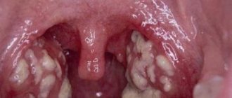
Cleft palate in children, photo
In children with this problem, inflammation of the middle ear is more often observed, as a result of which the development of hearing loss is likely.
Since food often enters the nasal cavity due to this pathology, chronic inflammation of the nasal cavity often develops.
In such patients, the development of bones and cartilages of the facial skull is disrupted, and its deformation appears.
Due to the pathology, the child breathes shallowly, which leads to oxygen deficiency.
Cleft palate can be associated with other developmental disorders.
Thus, if a child is born with a cleft palate, he will experience the following symptoms:
- aesthetic pathology of the face;
- problems with eating due to the presence of a defect;
- speech disorder;
- other concomitant diseases provoked by this pathology.
Anatomy of the affected area
Let's take a brief look at what tissues are affected by a cleft lip. This will make it clearer the amount of work that the surgeon has to perform.
The lip is a complex musculocutaneous formation. The outer layer consists of three parts:
- skin, which contains mucous and sweat glands;
- the skin passes into the intermediate part, which has a slightly different structure and is rich in blood vessels (therefore has a red color);
- the intermediate part passes into the mucous membrane, which is in direct contact with the teeth.
Beneath the mucocutaneous outer layer is a loose layer of connective tissue, and below that is the orbicularis oris muscle and several other muscles. Below the lip are the gums, the mucous membrane that covers the bone of the upper and lower jaw.
The lower jaw is a solid bone. It consists of a body on which there are cells for teeth, and processes that connect to the skull. The upper jaw is more complex: it not only has cells for teeth, this bone continues higher and forms the entrance to the nasal cavity, as well as the maxillary sinus.
Tests and diagnostics
A doctor can diagnose this defect in a child even at the stage of intrauterine development by conducting an ultrasound examination.
After birth, the doctor, during the examination and through additional research, determines the shape of the lesion and the complexity of the pathology. The purpose of such studies is to determine the presence of possible developmental pathologies, hearing, breathing, and sound production disorders.
If this pathology is confirmed, additional consultation with specialists may be required: otolaryngologist, dentist, infectious disease specialist, allergist, etc.
In the process of diagnosing the condition of a baby with such a pathology, the following studies are carried out to determine his general health:
- Laboratory tests - general and biochemical blood tests, urine tests.
- ECG.
- Ultrasound examination of the abdominal organs.
- X-ray of the chest organs.
In children
Cleft palate in humans, photo
According to medical statistics, this pathology occurs in one out of 2,500 newborns. It is very important that this disease is diagnosed at the stage of intrauterine development, since a baby with cleft palate and lip may experience complications during childbirth: the likelihood of amniotic fluid entering the respiratory tract increases. The doctor leading the birth should know about this in advance.
After the birth of such a baby, it is necessary to provide him with appropriate care and medical attention, because children with such a defect are very susceptible to infections and are prone to developing a number of diseases.
It is important to provide such a baby with normal nutrition, because such children have difficulty latching on to the breast, and it is very difficult for them to suckle. But in many cases, thanks to patience and certain skills, breastfeeding can be established. If this does not happen, use a breast pump and bottle. In difficult cases, the child is fed through a tube.
Such a child should be fed in a semi-upright position to prevent oral contents from entering the respiratory tract. After the baby has eaten, he must be held in an upright position until a burp occurs. When the baby is lying down, his head should be turned to the side.
Cleft palate disease, photo
Sometimes, immediately after birth, the child is fitted with an orthopedic plate to facilitate feeding and help control the development of the upper jaw. The condition of the plate is regularly monitored before surgery.
Children born with this pathology have normal hearing. But due to the tendency to develop otitis media, hearing may subsequently worsen. Therefore, it is important to regularly visit an otolaryngologist and have your baby’s hearing checked.
As for speech defects, after surgical intervention, children with such problems are able to completely restore articulation and speech skills.
It is very important to monitor your baby's oral hygiene. As the baby's teeth grow, parents should gently brush them every day. It is very important to regularly visit the dentist, who will monitor the process of tooth growth and bite formation.
A child with such a pathology should be provided not only with proper care and monitoring of his physical condition, but also with psychological help. As children with clefts grow older, they may have difficulty adjusting to the reactions of others to their aesthetic defect. Children need to be helped not only to gain self-confidence, but also to learn not to be afraid of the hospital, doctors, or surgery. An experienced psychologist will help you cope with these problems.
How dangerous is the disease?
The defect, in addition to aesthetic problems, is accompanied by such phenomena as:
- Difficulty swallowing.
- Dental disorders . If the problem is not eliminated before the baby's first teeth begin to emerge, this may lead to the absence of some teeth, or, conversely, to the appearance of extra ones.
- Speech impairment . The child cannot pronounce certain sounds correctly, and his voice becomes nasal.
- Hearing impairment, possible otitis media.
- Difficulties in adaptation . The child experiences psychological problems related to his appearance.
Over time, the child’s bite becomes disturbed, and this is fraught with problems such as impaired digestion of food (since the child cannot chew it well), tooth sensitivity, and a tendency to form caries.
Diet
Diet Table No. 1b
- Efficacy: therapeutic effect after a week
- Terms: 10-30 days
- Cost of products: 1100-1200 rubles per week
Cleft palate defect, photo
Since breast milk is the ideal product for a newborn baby, it is important to do everything possible to maintain breastfeeding. If the baby cannot latch on to the breast, he should be fed from a spoon or from a special bottle, expressing milk.
A mother who feeds her baby with breast milk must adhere to a diet and not consume foods that provoke allergic reactions (red fruits and vegetables, sweets, red fish, etc.). If breast milk is not available, it is important to choose a suitable adapted formula. Your pediatrician will help with this.
As your baby grows, it is necessary to formulate a healthy diet for him. Sweets and fruit juices that contribute to the development of caries should be minimized, as children with cleft palate are prone to this disease.
After surgery, the child must adhere to a gentle diet (diet table No. 1b is used).
Consequences and complications
With proper treatment, in most cases the problem can be eliminated. After surgery, scars of varying severity may remain. But in general, the right approach to treatment allows you to get rid of a physical and aesthetic defect, and the person lives a full life.
Proof of this is the fact that many people who later became famous were born with this defect. For example, the famous actor Joaquin Phoenix was born with this pathology. Andrei Makarevich and Mikhail Boyarsky had a cleft lip at birth; the famous comedian Denis Dorokhov was born with this defect.
List of sources
- Vasilyeva E.P. Features of speech disorders in children with congenital cleft lip and palate // Children's Hospital. 2011. No. 1. P. 46-48.
- Gonchakova S.G., Gonchakov G.V., Prityko A.G. Tactics of surgical treatment of patients with recurrent defects of the hard palate // Dentistry 2004: collection. tr. 6 Russian Scientific Forum. M., 2004. pp. 41-42.
- I.A. Kozin “Aesthetic surgery of congenital facial clefts” - Moscow, 1996. “Martis” - 568 p.
- Kharkov L.V., Yakovenko L.N., Vyshpinsky I.M. Classification of congenital clefts of the upper lip and palate (Literature review) // Bulletin of Dentistry. 2009. No. 1. P. 107-115.