Contrary to popular belief, atheroma is not a tumor. This confusion arose due to several reasons, primarily due to the suffix characteristic of neoplasms (lipoma, hemangioma, fibroids, etc.). By its nature, this is a non-inflammatory disease of the sebaceous gland, in which a cyst is formed.
Atheroma can be located on almost any part of the body, but it occurs much more often in areas with “oily” skin - on the face, behind the ears, on the back of the head, in the projection of the forehead, on the back between the shoulder blades, on the tailbone, etc. This is explained by the increased number of sebaceous glands in these areas. We will talk about the treatment of atheroma.
General information
Atheroma - what kind of formation is this? Atheroma (trichodermal cyst or steatoma) refers to fairly common tumor-like neoplasms of the pilosebaceous apparatus of the skin. It is one of the varieties of the group of epithelial skin cysts and is a rounded encapsulated neoplasm filled with thick yellowish-white contents, which may have an unpleasant odor. The contents of the tumor are represented by the protein keratin , produced by the walls of the capsule and atheromatous masses. The atheroma capsule is represented by connective tissue and is lined with several layers of flat epithelial cells (Fig. below).
ICD-10 code: D23. Other benign skin neoplasms. Atheromas occur in 7–10% of the population. At the same time, atheromas occur more often in women compared to men, and in people of the older age group more often than in people under 30 years of age. Often found in patients with acne and seborrhea . It occurs predominantly in areas of the skin with a high concentration of sebaceous glands (scalp, breast, face, back), therefore it is often found in the literature as a sebaceous gland cyst. In most cases, the size of atheromas is 1-3 cm, less often they reach 5 cm, but in practice there are cases of large atheroma formations. The localization of atheromas may vary.
The most common are atheroma of the scalp and atheroma on the back in different places, as well as atheroma on the face (usually on the cheek and skin of the forehead, in the chin area). Less common are atheroma behind the ear, on the ears (atheroma of the earlobe or pinna), on the neck (lateral/back surface), and atheroma of the mammary gland. And much less often - on the leg, atheroma on the penis, on the scrotum or atheroma on the labia and on the skin in the groin in women.
Below are photos of sebaceous gland cysts of various locations:
Photo. Atheroma on the back
Photo of atheroma on the head
Photo of atheroma behind the ear
Atheroma on the leg photo
Atheroma (blockage of the sebaceous gland on the eyelid). Photo.
Atheroma and lipoma, differences
Often in everyday life, atheroma is called a wen . However, this is not true, since there is a fundamental difference between them. Externally, atheroma is certainly similar to lipoma , however, structurally they are fundamentally different. Lipoma consists of altered cells of adipose tissue and develops in the layer of subcutaneous connective tissue, while atheroma is a cyst of the excretory duct of the sebaceous gland.
Depending on the histomorphological structure, the sebaceous gland cyst of the skin is divided into several types:
- Epidermal cyst - occurs very rarely, is formed from the skin epithelium, which, due to disturbances in the embryonic period, was subdermally transferred, that is, submersible growth of epithelial elements occurs, their proliferation and desquamation, which leads to the formation of a cyst cavity, which is filled with keratin and components of the skin lard This type of atheroma is characterized by slower growth, occurs predominantly in females, the predominant localization is the scalp (atheroma on the head) and the perineal area (in the groin in women, atheroma on the penis).
- Retention - formed due to blockage of the excretory duct of the gland. It is characterized by faster growth and occurs in both sexes with equal frequency; in addition to the scalp, it can occur on the skin of the face, back, mammary glands, and external genitalia. They can be single or multiple, often of the same size. They tend to become inflamed and coalesce into lumpy conglomerates.
Atheromas are a pronounced cosmetic defect, which causes psychological discomfort and becomes the main reason for contacting a surgeon. Also, if an infection enters the internal cavity of the atheroma, there is a high probability of its suppuration, since the contents of the tumor are a favorable environment for the proliferation of bacterial microflora with the development of the inflammatory process and the formation of an abscess (cavity with pus).
Suppurating atheroma (there is no code according to ICD-10), since the atheroma code according to ICD-10: D23 does not provide for cyst complications. It often develops after mechanical damage to the atheroma, but in most cases the cause of its occurrence cannot be determined and therefore a diagnosis of “idiopathic purulent atheroma” is made.
Answers to frequently asked questions
- Is there a risk of atheroma reappearing?
Yes, even a small number of surviving cells can give rise to the appearance of a new cyst.
- What does a postoperative scar look like? Is it possible to avoid it?
Minimal scars are formed after surgery using radio wave radiation. In second place is the argon plasma method. After these interventions, there may be no scars at all. Traditional surgery leaves noticeable scars.
- How to prevent atheroma?
There are no specific methods of prevention. It is important to control hormonal levels, maintain hygiene, and avoid skin injury.
- What is the difference between atheroma and lipoma?
Lipoma is located in the thickness of the fatty tissue, it is caused by excessive growth of connective tissue and is a benign tumor.
- Can the process become malignant?
No, such cases are excluded, this is not a precancerous disease.
- Can cyst destruction occur on its own?
No, this is impossible, atheroma cannot resolve, it remains unchanged for a long time.
- Is a surgeon required to operate on non-inflammatory atheroma?
Services under the compulsory medical insurance policy may not provide for the removal of non-inflammatory atheroma. The solution is to undergo surgery in a private clinic or wait for inflammation to develop, which does not guarantee the absence of a cosmetic defect after the intervention.
- What are the consequences of self-squeezing atheroma?
The cyst is usually located in areas of intense blood circulation, so there is a high risk of infection spreading to the brain through the blood vessels if bacteria gets into the wound. You cannot squeeze out the cyst - you need to seek qualified medical help.
Author of the article:
Volkov Dmitry Sergeevich |
Ph.D. surgeon, phlebologist Education: Moscow State Medical and Dental University (1996). In 2003, he received a diploma from the educational and scientific medical center for the administration of the President of the Russian Federation. Our authors
Pathogenesis
The formation of atheroma occurs as a result of the cessation/impairment of the release of the secretion of the sebaceous gland. As the secretion accumulates, it expands the duct of the sebaceous gland, forming a cavity in it with contents of a dough-like consistency, which includes particles of fat, detritus (a product of the breakdown of necrotic tissue) and keratinized cells of the epidermis. The skin above the gradually enlarging cyst rises, and a round-shaped compaction with a soft-elastic consistency is formed. As it grows, a capsule of connective tissue begins to form around the overstretched walls of the sebaceous gland. At the same time, the inner surface of the cyst produces secretion. If an infection penetrates into the atheroma cavity, then a purulent atheroma develops, that is, an inflammatory process develops, and the contents of the tumor turn into a purulent mass (Fig. below).
Postoperative period
In the first days after removal of the capsule, it is important to carefully monitor the condition of the wound. At first, the surgeon performs daily procedures, changing the drainage device and antiseptic dressings. The total duration of the postoperative period is 10-14 days. It takes place on an outpatient basis. Occasionally, complicated forms of atheroma are treated in a surgical hospital.
After the formation of connective tissue between the edges of the wound, the sutures are removed. This manipulation takes no more than 3-5 minutes and is usually painless.
Classification
Depending on the origin there are:
- Congenital atheromas (primary, true) are a hereditary disease and develop from detached epidermal cells during the embryonic development of the fetus.
- Acquired (secondary, false) - are formed due to blockage of the sebaceous gland duct or difficulty in the outflow of its secretion, which contributes to the accumulation of secretion in the lumen of the gland and the formation of a sac filled with atheromatous masses (altered sebum).
How to recover after surgery?
The rehabilitation period will largely depend on the method used to remove the cyst. However, there are also general recommendations, following which you can return to normal as quickly as possible.
Step 1. Bed rest is recommended.
Maintain bed rest
Step 2. It is required to limit any physical activity and eliminate heavy lifting for at least a month.
Avoid physical activity
Step 3: It is important to stay warm and protect your body from exposure to cold temperatures.
Avoid the cold
Step 4. It is required to change the position of the body as often as possible, and for the first 3 weeks you cannot lie on your back or sit for a long time.
Change your body position regularly
Step 5. When the stitches are removed, you need to regularly wash the intergluteal fold with an antiseptic drug.
Rinse the intergluteal fold
Step 6. For the first six months after surgery, it is necessary to remove hair in the area of the intergluteal fold.
Remove hair within 6 months
Step 7. It is important to visit your doctor regularly for examinations and assessment of your general condition.
Visit your doctor regularly
If you want to learn in more detail how to treat a cyst on the coccyx in men, and also consider alternative treatment methods and rehabilitation, you can read an article about this on our portal.
Video - Pilonidal cyst
A coccyx cyst is a disease that is not to be trifled with. It is important to see a doctor in time and not delay treatment in order to avoid the development of serious complications. Self-medication should be completely excluded. And you can prevent the development of cysts only if you take good care of your health and follow the rules of personal hygiene.
Causes
The main causes of atheroma:
- Genetic predisposition.
- Metabolic disorders .
- Hormonal imbalance (changes in the ratio of testosterone and estrogen during pregnancy/menopause, during puberty, taking hormonal medications for a long time).
- Stress (sebaceous glands are activated in stressful situations and sebum production increases).
- Tendency to sweat.
- Failure to maintain personal hygiene, use of inappropriate decorative cosmetics.
- Abuse/improper implementation of cosmetic procedures with an aggressive effect on the epidermis.
- Damage to glands when squeezing out blackheads/pimples.
- Violation of the microbial composition of the skin.
- Inflammatory skin changes ( rosacea , acne , etc.).
- Unfavorable influence of the external/production environment.
These causes are risk factors for the formation of atheroma, but pathology develops only with prolonged exposure.
Why does it appear?
Under the influence of various factors, the duct can become clogged. Continued secretion leads to the accumulation of contents and a gradual increase in size of the gland. It becomes larger, sometimes reaching the size of a walnut and even a chicken egg, its walls become denser, and a “bump” is formed, which becomes clearly visible and dense to the touch. Its contents resemble crushed lard.
The main reasons for the appearance:
- trauma (bruise, abrasions, minor abrasions)
- neglect of hygiene
- congenital narrowness of the ducts
- violation of secret exchange
Symptoms
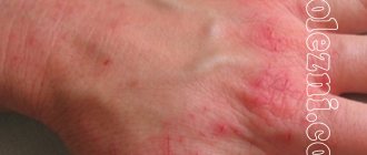
The standard complaint of patients is the presence of a superficial tumor-like neoplasm with a dense elastic consistency, often easily movable when pressed with a finger and painless when palpated. In uncomplicated cases, the skin over the cyst remains unchanged; less often, on the most elevated part of the skin above the cyst, an enlarged obstructed duct of the sebaceous gland is noticeable. With rapid growth of the tumor, the skin may ulcerate. In the case of a secondary infection and the development of inflammation, redness of the skin over the atheroma is determined (a photo of blockage of the sebaceous glands with inflammation is shown below).
Purulent atheroma of the back
In this case, there is severe pain during palpation and the patient’s general condition suffers (weakness, general malaise, increased body temperature). If the outcome is favorable, the pus breaks through the capsule or comes out through the ducts of the sebaceous glands. However, in some cases, pus can escape into the subcutaneous layer with the development of an abscess.
People's Councils
Considering the reviews and opinions of most people, treatment with herbs and improvised means can provide significant relief for various ailments, including skin tumors. It is worth noting that popular recommendations are not able to completely get rid of the disease, but will only help prevent suppuration.
Aloe juice
Grate 2 large leaves of agave and squeeze out the juice. Wipe the sore spot daily (make fresh juice each time). Has an effective anti-inflammatory effect.
Lamb fat
Add a little sunflower oil and garlic juice to the melted lamb fat. Mix everything and put it in a convenient jar. Rub daily into the sore spot.
Burdock with butter
Grind the burdock root, add butter, keep in a dark place for three days. The medicine is ready for external use.
Egg film
Carefully separate the film from the shell and apply it to the site of the disease. Despite redness or swelling, continue the procedure at least 3 times. As a result, the pus will come out.
Share with friends
Rate this article
Tests and diagnostics
Diagnosis of atheromas in the presence of characteristic clinical symptoms does not present any particular difficulties. If necessary, an ultrasound examination of soft tissues can be performed, which allows visualization of the cavity and capsule with curd contents. Additional histological diagnosis is carried out during surgery by taking atheroma tissue for histological examination. Differential diagnosis is carried out with other types of soft tissue tumors ( lipomas , lipomas , hygromas , dermoid cysts , osteomas , malignant neoplasms ).
Determining the diagnosis
A skin formation can be diagnosed by external examination; it has a round shape.
At first glance, it seems that diagnosing such a disease is not difficult. However, complexity exists, and it lies in the similarity of the characteristics of other formations discussed above.
To clarify the presence of the disease, ultrasound is used, and it is the main diagnostic method. If necessary, in particular with purulent inflammation, laboratory tests are recommended. A puncture is performed to determine the condition of the cyst contents.
An analysis of secretions is carried out to exclude such dangerous diseases as acrodermatitis caused by the action of the borreliosis spirochete,
Atheroma in children
Atheroma in a child is a fairly rare occurrence, since the sebaceous glands in young children still function poorly. If adult patients require surgical removal of atheroma, then in most cases children under three years of age do not undergo surgery at all. An operation to remove atheroma in a child is undesirable, since general anesthesia is required, which is harmful to a still fragile body. Local anesthesia is not effective, since complete immobility is required, and in the case of a small child, this condition is almost impossible to comply with. Surgery is performed if:
- There is rapid aggressive growth of the cyst.
- The surrounding tissues/vessels swell and are compressed.
- Signs of inflammation of the cyst appeared.
- The tumor is disturbing the child.
Treatment methods
Contrary to popular belief, the formation of a sebaceous gland does not always require any urgent treatment. Other patients live with atheroma for years or decades.
A puncture, or even more so, squeezing out, is not an option. The neoplasm will persist as long as there is a capsule.
Various situations may be the reason for medical intervention:
- the atheroma is inflamed, especially important if this is not the first time this has happened;
- the neoplasm demonstrates active growth dynamics or is already too large (more than 2 cm in diameter);
- the cyst is a serious cosmetic defect or causes pain.
Treatment in this case is a priori surgical. Conservative therapy is only additional during the healing stage.
Drug therapy
It is almost impossible to remove a cyst with cosmetics. Never the true form, but if you catch the secondary form at the very beginning of its formation, then there is a chance to prevent the formation of a fibrous capsule.
Products for cleansing the sebaceous glands will help here:
- Botanical Effects cleansing gel;
- Mary Kay cleanser;
- Clear proof gel for problem skin;
- special cleansing soap TimeWise.
The beginning of an infectious inflammatory process is well relieved by Dimexide, Levomekol and other external antibiotics.
Surgical methods
Surgery to remove atheroma is almost always performed under local anesthesia, since this surgical procedure is minimally invasive.
In traditional surgical practice, 2 methods are used:
- After miniature excision of the dermis with a special forceps, the surface of the skin is carefully shifted without damaging the capsule of the neoplasm. — By pressing the edges of the wound with your fingers, the cyst is removed.
- The second method involves removing large atheromas, and not just small and medium ones. - Here you will need the help of special tools. — Two bordering incisions are made on top of the cyst, into which the jaws of special curved surgical scissors are inserted. - This is done in order to bring them under the bottom of the neoplasm. This is how husking is carried out. — This method is excellent for removing pathology on the head, since the cosmetic defect with this method remains very insignificant.
Simply opening such a pathological structure is useless and extremely painful for the patient.
Laser removal
Laser removal of atheroma has a number of advantages:
- painlessness;
- absence of cosmetic defects;
- guarantee of protection against relapse;
- general minimally invasive procedure and high speed of its implementation;
- The patient can go home immediately after the procedure.
The atheroma is carefully opened, then the contents are evaporated with a high-intensity laser beam.
The disadvantages include:
- the high cost of such treatment (not included in compulsory medical insurance);
- Only small and medium cysts can be removed with laser.
And not every domestic clinic has the appropriate equipment.
Radio wave excision
Radio wave removal of atheroma is based on the fact that high-frequency radio wave radiation enhances the movement of molecules in organic tissue. Which leads to the release of heat, and accordingly, the destruction of this tissue.
The radio wave method, like the laser method, has similar advantages:
- low degree of trauma;
- speed of execution;
- comfort for the patient and almost complete absence of traces of the operation;
- fast regeneration;
- there is no risk of burns or tissue necrosis.
But the disadvantages are the same:
- high price;
- the use of the method is not possible in every clinic;
- not suitable for large tumors.
Radio wave surgery is especially helpful during operations on the face.
Electrosurgery
Electrosurgery is based on the principle of converting a high-frequency electromagnetic field into thermal energy, which destroys diseased or atypical tissue.
This method is used within the same framework as the previous two.
Home medicine
Treating atheroma at home is pointless and sometimes downright dangerous to health.
Folk remedies help the healing process after cyst removal. In the same way, they fight problematic skin that is prone to purulent acne, against which there is a risk of secondary atheroma.
Home remedies to help the skin with inflammation of atheroma:
- Vishnevsky ointment. — Proven by numerous practices, a product based on birch tar. — It has a pronounced antibacterial effect and is distinguished by the fact that it “pulls out” purulent masses from the affected tissue. “With the help of this liniment you can quickly relieve inflammation, but Vishnevsky ointment is not suitable, and is not even acceptable for open bleeding ulcers.
- Hydrogen peroxide and iodine. - There is no need for introductions, because these means are familiar to everyone. — They have an antiseptic and anti-inflammatory effect.
- Ichthyol ointment. — Another antiseptic external agent based on ammonium salt, sulfonic acids and shale oil. — It is considered a safer remedy than Vishnevsky’s liniment.
It should be remembered that certain patients may be allergic to some traditional recipes.
Traditional healers claim that it is possible to cure atheroma without surgery. But there is no reliable scientific evidence for this.
Postoperative care
The recovery period depends on the following factors:
- tumor size;
- method of surgical intervention;
- pathology scenario (with complications, without complications);
- quality of healing and postoperative recovery.
You can wash your hair after removing a small atheroma literally the next day. Especially if the operation was performed using some new minimally invasive method.
If dressings are prescribed after removal of atheroma, do not change the dressing yourself.
It is not recommended to exercise immediately after surgery, even if the tumor was small. Physical activity causes active sweating, which can trigger cyst recurrence.
If everything goes without complications, then the patient can return to their usual rhythm of life a week after the procedure.
When small formations are removed, the scar resolves on its own. However, even with the formation of a keloid scar, this cosmetic defect is easily eliminated within the framework of the same laser surgery.
Consequences and complications
A frequent complication is suppuration of atheroma. Infection of the cyst leads to the development of an inflammatory reaction, hyperthermia , and disruption of the general condition. Of particular danger is abscessed atheroma and cases when they try to squeeze out its contents, which contributes to the spread of pyogenic microorganisms in the tissues and leads to phlegmon , and in cases where they enter the general bloodstream, to sepsis . Complications include relapse of atheroma (re-formation of a cyst) after poor-quality removal or its spontaneous opening. Cosmetic complications include the formation of colloidal scars after tumor elimination. In extremely rare cases, epidermoid atheroma can transform into a malignant tumor. Malignancy of retention atheromas is impossible.
What is a neoplasm?
Such benign formation is divided into:
- true (primary);
- false (secondary).
A sebaceous cyst of true etiology is formed during embryogenesis, for example, as a result of teratogenic effects. It is often located under the skin in an undeveloped state, without bothering the patient during his life.
But according to statistics, patients with a false form seek medical help.
In this case, what happens:
- suppuration of atheroma, a focus of inflammation is formed, sometimes so powerful that it leads to general hyperthermia and intoxication;
- a fibrous capsule is formed;
- periodic breakthrough of the cyst with the formation of an ulcer, a crust forms on the affected tissues;
- in rare cases, malignancy (malignancy) is possible.
The content of atheroma is a sebaceous secretion (sebum), which differs in physical and chemical characteristics from ordinary fatty secretions of the skin.
False atheromas are a consequence of other systemic disorders in the body. With small sizes (up to 5 mm), it is capable of passing on its own with a moderate probability.
True atheroma, if it has arisen, will not disappear without intervention.
List of sources
- Kapustina O.G. Diagnosis and optimization of treatment of skin tumors in the outpatient practice of a dermatologist. dis. Ph.D. honey. Sci. – M., 2009. – 163 p.
- Dubensky, V.V. Skin neoplasms in the practice of a dermatovenerologist Text. / Edited by V.V. Dubensky // Tver: “Publishing house-Triad”. 2002. - 148 p.
- Bezrukov S.G., Grigorieva T.S. Histomorphological features of the structure of the membrane of facial atheroma // Tauride medical and biological bulletin. - 2013. - T. 16, No. 1, part 3 - P. 37-41.
- Kubanova A. A., Kubanov A. A., Rakhmatulina M. R., Malova I. O., Sokolovsky E. V., Apolikhina I. A., Melkumyan A. G. Federal clinical guidelines for dermatovenereology. Dermatovenereology 2015: Skin diseases. Sexually transmitted infections. M.: Business Express, 2021. 768 p.
Spinal cord cyst
A spinal cord cyst is a cavity filled with fluid. It is formed in the spinal cord trunk, in the spinal cord itself. Cystic formation appears due to severe injuries, against the background of various diseases and infections. Most often, cervical spinal cord atheroma is detected.
Important! Sometimes a spinal cyst is a congenital pathology that appears against the background of genetic abnormalities. Education begins to increase in adolescence or early 20s.
This formation is very dangerous for health, since it compresses the spinal cord, increasing in size. This leads to a lack of coordination. If a spinal cord cyst is diagnosed, the atheroma hurts, weakness in the legs occurs, and sensitivity decreases. Legs and arms go numb, muscle weakness increases.