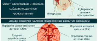
Aortic aneurysm is a pathological expansion of the largest vessel in the human body, which is accompanied by thinning of its wall with the likelihood of rupture and fatal bleeding. Thrombotic masses form in the aneurysmal sac, which can be washed away by the blood flow and cause blockage of the arteries of the lower extremities or internal organs, which can lead to the development of acute arterial insufficiency and gangrene. Complications of aortic aneurysm account for 6% of all deaths in developed countries. However, there is an effective and safe treatment that reduces the risk of such complications tenfold.
Different types of pathology
Aortic pathology is common among older people. It is extremely rare in women, which cannot be said about the stronger half of humanity. Pathology can develop for a very long time, for years. The patient needs regular care and medical supervision. Lifestyle plays a huge role.
Aortic pathology can be classified according to etiology, shape, segments and wall structure. Based on this, it is divided into subspecies, each of which has its own characteristics and manifestations. Aneurysms are distinguished by segments:
- aortic arch;
- sinus of Valsalva;
- ascending department;
- descending department;
- abdominal aorta.
In addition, an aneurysm can be combined, that is, it affects several areas at once. In this case, special treatment is needed, step by step.
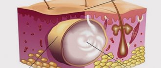
Morphological differences in aortic disease divide it into false and true. In the latter case, the membrane thins and bulges outward. This happens with atherosclerosis, syphilis and similar diseases. In the false one, hematomas are detected. Appear after interventions by a surgeon or as a result of injury to an organ. This is quite possible as a consequence of surgery on the organ.
According to the shape, the pathology of the aorta is divided into saccular and fusiform. In the first case, there is a bulging of the walls outward, locally. In the second, the same thing happens, but over the entire diameter of the aorta. Depending on how the disease progresses, it can be:
- uncomplicated;
- complicated;
- exfoliating.
The most serious is complicated. It often leads to rupture of the aortic sac. As a result, internal bleeding, hematomas, and thromboembolism are observed. As a consequence, death is obvious, and almost instantaneous due to blood loss. If there are no qualified medical professionals nearby, this aortic problem cannot be dealt with. It is for this reason that the patient should always be under medical supervision.
Internal iliac artery
It descends to the psoas major muscle, namely to its medial edge, and then runs down, penetrating into the small pelvis. In the area where the sciatic foramen is located, the artery is divided into a posterior and anterior trunk. The latter are responsible for the blood supply to the tissues of the walls and pelvic organs.
The internal iliac artery has the following branches:
- iliopsoas;
- umbilical;
- upper, lower gluteal;
- middle rectal;
- lower vesical;
- internal genitalia;
- obturator;
- uterine.
In addition to the listed branches, this artery also gives off parietal and visceral branches.
What causes the disease to develop?
Regardless of the form, aortic pathology can be acquired or congenital. Congenital aortic aneurysm is formed due to diseases that are often transmitted at the genetic level from relatives. These include fibrous dysplasia, hereditary elastin deficiency and other syndromes. If the disease is acquired, the causes may be arthritis, infections or fungal infections. But pathology can occur without an inflammatory process, for example, as a result of atherosclerosis, prosthetic defects and suture material.
Mechanical causes are common. In this case, it means both external and internal damage to the organ. This happens due to an incorrectly performed surgical operation on an organ or after it.
Known causative factors that increase risks are:
- advanced age;
- alcohol;
- smoking.
More often, pathology is detected among representatives of the stronger sex. Aneurysm of the aortic arch and its other locations often manifests itself as a result of defects in suture materials and grafts. Simply put, after various operational actions. Post-traumatic consequences are not uncommon today. After injury, pathology does not appear immediately: it can take from a month to several years. There is evidence of cases where aortic disease made itself felt after 20 years.
Hypertension weakens the body's tone, which creates an aneurysmal sac. This mainly happens after 60 years. Increasing blood flow pressure only increases the risk. This becomes the result of internal bleeding, which in turn has tragic consequences. To prevent this from happening, you need to know the symptoms of an aneurysm.
Iliac artery
The iliac artery is the largest paired blood vessel after the aorta, five to seven centimeters long and 11-13 mm in diameter. The arteries begin at the bifurcation of the aorta, at the level of the fourth lumbar vertebra. In the area of the articulation of the iliac bones and the sacrum, they break up into the external and internal iliac arteries.
The internal artery splits into branches - middle rectal, iliopsoas, sacral, lateral, inferior and superior gluteal, inferior vesical, internal genital, obturator. They deliver blood to the organs and inner walls of the pelvic cavity.
The external artery, leaving the pelvic cavity, simultaneously gives off several branches to its walls and continues in the area of the lower extremities in the form of the femoral artery. The branches of the femoral artery (profunda artery, inferior epigastric artery) deliver blood to the skin and muscles of the thighs, and then branch into smaller arteries and provide blood supply to the foot and leg.
In men, the iliac artery delivers blood to the membranes of the testicle, thigh muscles, bladder, and penis.
Symptoms of pathology
Any aortic aneurysm is identified based on its characteristics, depending on its location, length, size and other factors. In some cases, it does not show obvious signs. It is discovered accidentally during routine examinations. If there are symptoms, then the main symptom is always the same - pain arising from stretching of the aortic lining.
With abdominal disease, the following symptoms can be observed:
- Painful sensations.
- Discomfort in the abdominal area.
- Heaviness.
- Belching.
- Feeling of full stomach.
With an aneurysm of the ascending section, the following appear:
- Heart pain.
- Dyspnea.
- Tachycardia.
- Dizziness.
If the pathology of the aorta reaches a large size, headaches, swelling of the chest and face may occur. This happens due to the pressure of the expanding aorta on neighboring tissues. In this case, immediate medical attention is required, as in all other cases.
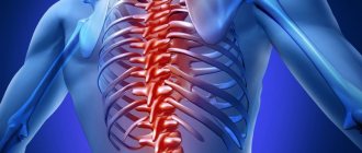
When the descending aorta of the heart is irritated, pain appears in the shoulder blade and arm, on the left side. Often the pain radiates to other areas of the body. Spinal cord ischemia and paraplegia are possible.
When the aortic arch is damaged, compression of the esophagus is observed, as well as:
- dysphonia;
- bradycardia;
- dry cough;
- salivation;
- dyspnea.
The greater the pathology of the aorta becomes, the more it compresses neighboring anatomical structures - nerve plexuses, tissues. In this case, there is often pain behind the chest, throbbing, pain radiating to the shoulder, neck and back. Horner's syndrome appears, and the pupils become constricted. It is by these symptoms that you can promptly identify the pathology yourself.
Disease prognosis
As for the prognosis of the disease, it largely depends on the size of the aneurysm, on the presence of atherosclerosis, on the location and type of defect. If we consider the prognosis as a whole, it remains unfavorable, which is associated with a high risk of death of the patient from aneurysm rupture. If the formation reaches 6 or more centimeters, then the probability that it will rupture within a year is 50%, and with an aneurysm of a smaller diameter, the percentage of its rupture is reduced to 20%.
The earlier an aortic aneurysm is detected, the more favorable the prognosis that the operation will be successful and the lower the mortality rate of patients during its implementation.
Author of the article:
Molchanov Sergey Nikolaevich |
Cardiologist Education: Diploma in Cardiology received from Perm State Medical University named after. I. M. Sechenova (2015). Here I completed my postgraduate studies and received a diploma in Cardiology. Our authors
How is an aortic aneurysm diagnosed?
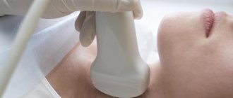
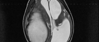
A number of diagnostic measures are used to detect aortic aneurysms. X-rays, tomography and ultrasound examinations are performed. Systolic murmurs are detected in the aorta. However, diagnosis begins with palpation. It reveals a pulsating swelling, indicating the presence of an aneurysm. External examination is the basis of diagnosis. In addition to pulsation, it helps to identify protrusions of the aortic sac. An anamnesis is taken to identify secondary diseases or injuries. This will help confirm or refute the presence of pathology.
After manual study, instrumental study must be performed. It begins with radiographic studies. Diagnosis includes plain radiography of the abdominal cavity, fluoroscopy, radiography of the stomach, esophagus and chest. It detects ECG deviations well; an ultrasound scan can also be prescribed. A CT scan of the abdominal or thoracic aorta identifies possible dilations of the arteries, blood clots, and hematomas.
Finally, aortography is performed to determine the localization of the pathology, its extent and size. Only such comprehensive diagnostic actions make it possible to establish an accurate diagnosis and develop appropriate treatment. After this, you can begin to implement therapeutic procedures.
Classification
By etiology: congenital and acquired (atherosclerotic, traumatic, syphilitic, mycotic, due to nonspecific aortitis)
Shape: spindle-shaped, bag-shaped
By type (prevalence) of damage:
Type I - damage to the proximal segment of the abdominal aorta with involvement of the visceral branches;
Type II - damage to the infrarenal segment before the bifurcation;
III - infrarenal segment involving the bifurcation and iliac arteries;
IV - total damage to the aorta.
Localization (direction) of the rupture: retroperitoneal space, free abdominal cavity, rare (inferior vena cava, duodenum, bladder, etc.).
Troubleshooting
When an aortic aneurysm is confirmed, it needs to be repaired. If the pathology does not show visible symptoms, then dynamic medical observation is sufficient. In this case, regular X-ray examinations play an important role. Of course, procedures are carried out in parallel to prevent complications using different therapy methods. Medications play an important role here.
If the aneurysm reaches a large size, then surgery cannot be avoided. If the pathology progresses intensively, surgical treatment is also necessary. Emergency measures are needed for ruptures. In all such situations, the main measure can be considered excision of a section of the vascular system. It is possible to replace the defective area with a prosthesis or stitch it together. In general, two methods can be used - surgical and medicinal. But it all starts with therapy, that is, conservative prevention is carried out.
Modern treatment of aneurysm
Complications of an aneurysm require emergency surgery. Timely surgical intervention can save life. Such an intervention should be carried out in a hospital that has everything necessary for both endovascular and open surgical treatment of an aneurysm. Although a ruptured aneurysm can be repaired, only about 50% of patients are saved in these cases. Even if the patient survives the operation, he will often develop kidney complications, intestinal necrosis, or leg ischemia. Any other complications associated with aneurysms, such as embolism, abdominal pain, intestinal obstruction, require urgent surgery to eliminate it. Planned treatment has certain indications, which depend on the location of the aneurysm, the patient’s age, and concomitant diseases of the heart and lungs. If the aneurysm diameter exceeds 50 mm, the risk of rupture becomes unacceptable. Abdominal pain, embolism in a limb, and intestinal dysfunction due to an aneurysm are indications for surgery. If the aneurysm increases in size by more than 10% per year, this is also a risk factor for rupture and forces a decision on surgical treatment. The risk of rupture of an abdominal aortic aneurysm with a diameter of 7 cm or more is almost 20% per year. There is no easy method to repair an aortic aneurysm. This pathology poses serious difficulties for surgeons.
Conservative methods
For isolated aneurysms, this approach is justified if the lesion is small in diameter or symptoms do not appear. Various herbal formulations and tablets are prescribed:
- statins;
- antihypertensive drugs;
- adrenergic blockers.
When carrying out such recovery, dynamic observation is important. In this case, the affected organ is regularly examined by a cardiologist. MRI, CT, Echo CG are prescribed.
The main goal of drugs used in conservative treatment is to relieve symptoms when they are detected. Reducing risk and preventing the growth of pathology are also important goals of the technique. In addition, this is a kind of prevention, and very effective. At the same time, you need to understand that not a single medicine is capable of completely getting rid of the pathology, but only pushes it aside and freezes it. To ensure that the aneurysm no longer bothers you, radical techniques are required.
This treatment of the root of an aneurysm should be performed under the direction of an experienced professional with medical training. Self-medication will not give positive results, but it may well cause harm. Therefore, it is extremely important to take only those medications prescribed by your doctor. Otherwise, death is possible.
Internal iliac artery
It descends to the psoas major muscle, namely to its medial edge, and then runs down, penetrating into the small pelvis. In the area where the sciatic foramen is located, the artery is divided into a posterior and anterior trunk. The latter are responsible for the blood supply to the tissues of the walls and pelvic organs.
The internal iliac artery has the following branches:
- iliopsoas;
- umbilical;
- upper, lower gluteal;
- middle rectal;
- lower vesical;
- internal genitalia;
- obturator;
- uterine.
Surgical techniques
Such treatment is carried out when an aneurysm larger than 5 cm in diameter is detected, if there is compression syndrome, pain, dissection and other complications, such as thrombosis. This technology consists of resection. With its help, the aneurysm is dissected. The aortic defect is eliminated by replacing the affected area with a graft. This method is the most common. Of course, such an operation is very complicated, but almost always it guarantees complete relief from the pathology.
This procedure is carried out only after starting artificial blood flow. It is worth mentioning that such surgery sometimes ends in death. Therefore, the selection of a clinic and medical personnel for its implementation must be approached with special care. But of course, this is not the only method. Closed prosthetics are also used. In such a situation, an endoprosthesis is used. It is inserted into the lumen of the aorta, where it is fixed below or above the aneurysm sac.
There are cases when carrying out any of the operations described above is unacceptable. These include identifying complete contraindications. In this case, the affected artery is wrapped in synthetic fabric. Such palliative intervention is relevant only when there is a threat of rupture. In other cases, the patient's stable condition is coordinated by regular medications.
Patient rehabilitation
The duration of recovery depends on the type of intervention. The early stage of all operations is carried out in the intensive care unit and/or intensive care unit. In the postoperative period, intestinal problems are often observed.
After open surgery, the time spent in hospital is up to 14 days. Before discharge, the patient undergoes laboratory, ultrasound, and x-ray examinations. The patient can return to work after 10 weeks.
With minimally invasive intervention, the rehabilitation period is much shorter.
Modification of life after discharge from hospital:
- quitting smoking and alcohol;
- fractional healthy nutrition;
- a calm, stress-free life – especially in the first month;
- Contact, dynamic and power sports are prohibited;
- It is prohibited to lift weights over five kg at home;
- body weight control;
- blood pressure control at 130/85 mm Hg.
After six months, intensive walking, swimming, and then light running are allowed.
After such surgical interventions, the patient can be sent for recovery to a cardiological sanatorium. Such rehabilitation is indicated no earlier than the fourteenth day after surgery, if the patient’s condition is satisfactory and there are no complications.
Permitted spa treatment methods:
- phytotherapy;
- barotherapy;
- balneotherapy;
- exercise therapy;
- hydrogen sulfide baths;
- mineral water.
Possible complications
If, when aortic disease is detected or a pathology is suspected, serious treatment is not carried out, death is inevitable. This happens due to a number of consequences. With this pathology, the most dangerous thing is rupture of the aortic aneurysm, leading to serious bleeding. Shocks and collapses, heart failure are possible. When ruptured, conditions often transform, leading to death. These include:
- cardiac tamponade;
- hemothorax;
- hemopericardium.
If blood clots form in the aortas, when they break off, acute occlusion, soreness of the fingers, cyanosis, and intermittent claudication may develop. A stroke is also possible.
Most often, aortic defects and heart failure appear. Such complications are characteristic of pathologies in the ascending aorta. Especially if their origin is syphilitic. It is quite possible to develop decompensation of the heart. As mentioned, the most serious of them is rupture with bleeding. The flow of fluid from the veins can go into the bronchi, trachea, cardiac sac, pleural cavity, esophagus, even into large vessels of the chest. Thus, cardiac tamponade is more likely to occur. Rapid blood loss causes rapid death.
Another serious complication is blood clots in the aorta. Subacute and acute thrombosis occurs more often in the abdominal aortas. When they overlap, the most dire consequences can occur. As in other cases, this always leads to rapid death. Only measures taken in a timely manner will help. Accordingly, the patient should be under medical supervision at this moment. If all necessary measures are taken, an aneurysm will not cause problems.
Endovascular interventions
They allow you to dramatically reduce the amount of surgical trauma, shorten hospitalization periods and reduce the inevitable suffering of the patient associated with surgical approaches. One of the main disadvantages of the method is the need for repeated interventions.
Types of endovascular operations for aortic aneurysm:
- implantation of a stent graft into the abdominal aorta,
- implantation of a stent graft into the ascending (thoracic) aorta.
The most modern method of treating aortic aneurysm is a hybrid method, which allows achieving optimal treatment results with the least surgical trauma.
Hybrid surgeries combine the advantages of open and endovascular interventions.
To prevent the development of aortic aneurysms, the most important thing is the need to control risk factors, namely arterial hypertension. In addition to arterial hypertension, the most significant risk factors are age (over 55 years), male gender, smoking, the presence of aneurysms in direct relatives, and high cholesterol levels.