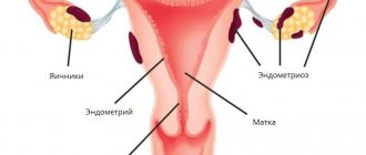
Uterine cancer is a malignant tumor that is localized in the body or cervix of the organ. Clinical symptoms of uterine cancer at an early stage may most often be absent or mild, so the disease is often diagnosed in the last, advanced stages, which are difficult to treat and often have an unfavorable prognosis.
Therefore, an important role belongs to timely diagnosis and detection of malignant neoplasms before the pathological process spreads to other organs. Every woman needs to know the first signs of uterine cancer at an early stage and, when they appear, immediately contact a gynecologist to clarify the diagnosis and immediately begin treatment.
What is uterine cancer?
This type of tumor is a neoplasm. As is known, neoplasms can be malignant or benign.
A tumor such as uterine cancer can be classified as a malignant tumor.
The formation of such a neoplasm originates, first of all, from tissues located in the uterus, which can spread to all parts of the body.
Oncological disease is one of the most common diseases and ranks fourth after cancer of the breast, skin and gastrointestinal tract.
Disease prognosis and follow-up
Malignant tumors of the uterine body in the initial stages can be classified as diseases with a relatively favorable prognosis. According to hospital registries, the overall 5-year survival rate of patients with uterine cancer who received treatment in the world's leading clinics reaches almost 80% (Figure 5).
Rice. 5. Overall 5-year survival rate of patients with endometrial cancer (Annual report on the Results of Treatment in Gynecological Cancer FIGO, 2006)
After completion of treatment, the patient needs follow-up visits to a gynecological oncologist and regular examinations to prevent the disease from returning.
Morbidity statistics
In order to talk about any cancer disease, of course, one cannot fail to note the statistical data on the basis of which appropriate conclusions can be drawn.
As mentioned earlier, uterine cancer is one of the ten most common cancers and ranks fifth among them.
Of course, it should be noted that the occurrence of this disease, as well as mortality from this pathology, has decreased significantly in recent years.
Statistics show that this pathology occurs more often in women over 50 years of age. However, according to doctors, young girls are also susceptible to this disease.
Previously, there was an opinion that uterine cancer is one of the main causes of death from malignant tumors. The incidence of such pathology has decreased to 70%.
How to detect cancer in the early stages?
Most endometrial tumors are detected in postmenopausal women. Therefore, the first symptom of uterine cancer is bleeding, which alarms most older women. In such cases, urgent consultation with a doctor and appropriate examination are necessary.
In young women, spotting is not so rare that a serious illness can be suspected. Therefore, it is very important to know the length of your menstrual cycle, the amount and duration of bleeding. If there are changes in the usual menstruation schedule, it is better to undergo an examination to exclude all serious causes.
Causes of uterine cancer
Of course, the formation of uterine cancer is promoted by certain causes and factors that can aggravate the severity of this serious disease.
As such, the exact reason why the development and growth of a neoplasm on the uterus begins in the modern world has not been established or studied.
Research has made it clear that the factors contributing to the growth of cancer include the following number of reasons:
- diabetes;
- smoking;
- hypertension;
- AIDS;
- human papillomavirus infection;
- failure of menstruation;
- venereal diseases;
- sexual activity at an early age;
- inability to have children;
- taking contraceptives;
- giving birth at too young an age.
One of the most basic and, perhaps, dangerous factors contributing to the formation of cancer is increased body weight.
If a female patient’s body weight exceeds the usual established norm by more than 10-25 kilograms, then the risk of developing a tumor will be tripled.
Some facts also play a very important role in the occurrence of a malignant tumor:
- erosion;
- ulcerative processes
- endometriosis of the uterus
- scar formations after childbirth;
- inflammatory processes.
Causes and risk factors
For many cancer pathologies, the exact cause of their occurrence is unknown. This also applies to uterine cancer. Pathology is considered a “disease of civilization” that occurs under the influence of unfavorable external conditions, dietary habits and lifestyle.
Factors predisposing to uterine cancer:
- late first menstruation;
- menopause only after 55 years;
- long-term anovulation;
- endocrine infertility;
- polycystic ovary syndrome and hormonally active tumor of these organs (Brenner cancer);
- obesity;
- diabetes;
- long-term use of estrogen hormones without combination with gestagens;
- treatment with antiestrogenic drugs (Tamoxifen);
- lack of sexual activity or pregnancy;
- cases of illness in close relatives.
Endometrial cancer of the uterus occurs against the background of a complex of disturbances in hormonal balance, metabolism of fats and carbohydrates.
The main pathogenetic types of the disease:
- hormonal-dependent (in 70% of patients);
- autonomous.
In the first option, ovulation disorders in combination with obesity or diabetes lead to increased production of estrogen. Acting on the inner uterine layer - the endometrium, estrogens cause increased proliferation of its cells and their hyperplasia - an increase in size and a change in properties. Gradually, hyperplasia becomes malignant, developing into precancer and uterine cancer.
Hormone-dependent uterine cancer is often combined with a tumor of the intestine, breast or ovary, as well as with ovarian sclerocystosis (Stein-Leventhal syndrome). This tumor grows slowly. It is sensitive to progestogens and has a relatively favorable course.
Signs that increase the risk of hormone-dependent cancer:
- infertility, late menopause, anovulatory bleeding;
- follicular ovarian cysts and hyperplastic processes in them (thecomatosis);
- obesity;
- improper treatment with estrogen, adrenal adenoma or cirrhosis of the liver, causing hormonal changes.
The autonomous variant often develops in postmenopausal women against the background of ovarian and endometrial atrophy. There is no hormonal dependence. The tumor is characterized by a malignant course, quickly spreading deep into the tissues and through the lymphatic vessels.
There is a genetic theory of cancer, according to which cell mutations are programmed into DNA.
The main stages of the formation of a malignant tumor of the uterus:
- lack of ovulation and increased estrogen levels under the influence of provoking factors;
- development of background processes – polyps and endometrial hyperplasia;
- precancerous disorders - atypia with hyperplasia of epithelial cells;
- preinvasive cancer that does not penetrate beyond the mucous membrane;
- minimal penetration into the myometrium;
- pronounced form.
Methods for diagnosing the disease
Diagnosis is a very important stage for any type of cancer. It is very important to diagnose the disease and this process must be organized competently.
Diagnostics includes:
- Routine doctor's examination.
- With the help of gynecological mirrors , the doctor is able to notice that there are any changes in appearance.
- In the future, the patient must be sent for an ultrasound , which will detect and determine the size and altered structure of the uterus. In addition, an ultrasound study determines the structurization and thickness of the endometrium.
- Curettage techniques and analysis of the histology of biological material are often used to determine the further fate and nature of the disease. This procedure is performed under general anesthesia in a hospital stay.
Degree of tumor differentiation
If a tumor is found, the doctor examines the tissue sample under a microscope to determine the type and grade of the tumor. This grade tells how different the type of cancer cells is from normal endometrial cells. The degree of tumor differentiation can determine how quickly it will grow. Highly differentiated tumors often grow faster and spread more often. The degree of differentiation also helps determine the further treatment protocol.
When diagnosing endometrial cancer, it is very important to determine the stage of the disease. This is the most important information for determining treatment tactics. The stages of the disease are determined based on whether the tumor has grown into neighboring organs or spread to other parts of the body.
When a tumor spreads to other organs, it consists of exactly the same pathological cells as the primary tumor and is called exactly the same. For example, endometrial cancer can spread to the lungs; in this case, endometrial tumor cells will be present in the lung. This is called metastatic endometrial cancer, not lung cancer. This complication is treated according to protocols for the treatment of endometrial cancer, not lung cancer.
To determine whether the tumor has spread, your doctor may order one of the following types of tests:
- Lab tests. A Pap test can show whether the tumor has spread to the cervix, and blood tests can reveal abnormal liver and kidney function. Your doctor may also order a test for CA-125, which is often elevated in various types of cancer.
- Chest X-ray: A chest X-ray can help identify a tumor in the lung.
- Computed tomography: Computed tomography takes many sequential X-ray slices, covering a large number of organs, and allows you to get a highly accurate picture and find a tumor in distant parts of the body: lymph nodes, lungs, etc. Sometimes computed tomography is performed with the introduction of a contrast agent.
- An MRI works like a large magnet that is connected to a computer and helps produce very accurate images of the uterus, other internal organs and lymph nodes. Sometimes an MRI is also performed with the injection of a contrast agent.
Often, surgery is necessary to accurately determine the stage of the disease. The surgeon removes the uterus and takes tissue samples from the pelvis and abdomen. When the uterus is removed, the specimen is examined to assess how deeply the tumor has grown. The surgeon also checks whether nearby organs and lymph nodes are damaged.
Symptoms of uterine cancer in women
Of course, symptoms play an important role in determining this disease.
A symptom is something that should be paid utmost attention to if the patient feels that something is wrong. It is extremely important for women over forty to pay special attention to their health.
Unfortunately, cancer is one of the diseases whose symptoms appear in the final stages.
Conventionally, symptoms can be divided into several types:
- The first stage is the signs and symptoms that appear before the onset of menopause. Let's assume that a woman is in the period of menopause. During such a period, the menstrual cycle gets confused and manifests itself in the form of discharge with blood, which becomes less and less in volume over time. During such a period, it is worth paying utmost attention to the discharge. That is, if previously they were rare, and later again abundant, this is the primary sign and symptom of the appearance of uterine cancer.
- There are also other signs during menopause . In the case when a woman has already reached menopause, and the menstrual cycle has not appeared for more than two months, symptoms of cancer can be any discharge with blood and the opening of bleeding.
Based on the age category and period of menopause, the following symptoms may appear:
- opening of bleeding;
- pain after intercourse;
- pain in the perineum;
- pain in the lumbar region and lower abdomen;
- fatigue and sudden weight loss.
If you have one of the symptoms, you must immediately visit a doctor to eliminate this problem.
Determination of uterine cancer before menopause
As noted earlier, there are symptoms that make it clear that a tumor has appeared before the onset of menopause.
Most often, during such a period, vaginal discharge is already irregular and appears less frequently with each passing month.
It is during this period that symptoms of uterine cancer can include all the discharge with blood from the vagina.
Uterine cancer can be suspected only if the menstrual cycle gradually stops, and then discharge in large quantities begins again.
Manifestation during menopause
At a time when a woman has already begun the period of menopause, namely menopause, symptoms may also arise that need to be given special attention.
As a rule, a woman has not had her period for several months; symptoms of cancer can include bloody discharge, regardless of the frequency with which they appear, for how long and in what volume.
Symptoms and signs
Uterine carcinoma does not have a specific clinical picture at the initial stages of development of the tumor process. This is the main reason why women seek medical help late. The presence of a neoplasm in the uterine cavity can be detected in a timely manner with regular examination. The initial signs of cancer are similar to other gynecological pathologies. Therefore, it is important to undergo a preventive gynecological examination in a timely manner. It allows you to detect cancer in the early stages. Only in this case does a favorable prognosis for recovery remain. The main clinical signs of the disease include:
- Bleeding from the uterine cavity;
- Beli;
- Pain syndrome;
- Purulent and watery discharge.
The appearance of uterine bleeding in women of non-reproductive age is the main sign of the presence of a malignant neoplasm.
Bleeding in women of reproductive age occurs in various gynecological pathologies. This does not always result in timely diagnosis of a uterine tumor. The appearance of uterine bleeding requires a mandatory examination to identify a malignant formation. Leucorrhoea is typical when the tumor increases in size. The amount of leucorrhoea varies and can be either scanty or abundant (leukorrhea). The accumulation of secretions causes aching pain in the lower abdomen. The nature of the discharge depends on the degree of tumor development. Leucorrhoea can be bloody, purulent, or bloody-purulent. This indicates the addition of an infection. This condition is accompanied by acute pain and intoxication syndrome. This indicates an extremely serious condition of the patient.
The severity of pain depends on the degree of tumor growth into surrounding tissues. In the initial stages of uterine cancer development, there is no pain syndrome. The appearance of pain occurs as the size of the cancer focus increases. The neoplasm can compress neighboring organs, causing associated clinical manifestations.
The accumulation of pus in the uterine cavity occurs with cervical stenosis. This manifestation of carcinoma is accompanied by symptoms of intoxication of the body. These include bursting pain in the lower abdomen, increased body temperature, chills, and sweating. Purulent accumulation in the uterine cavity requires immediate hospitalization and surgical intervention. The appearance of watery discharge is a specific sign of a uterine tumor.
Compression of the bladder by the tumor leads to frequent and painful urination. A large tumor volume can cause pain during defecation, as well as a painful urge to do so. In this case, excretion of feces occurs with difficulty. Compression of the ureter by an overgrown tumor leads to pain similar to the clinical manifestations of renal colic.
Against the background of intoxication, a decrease in appetite occurs. In this regard, body weight decreases sharply. An increase in body temperature occurs in the later stages of a uterine tumor. In this case, there is no effect from taking antipyretic drugs. Hyperthermia may indicate a secondary infection. Patients are also worried about weakness, sudden loss of strength, drowsiness, headache, dizziness.
The absence of distinctive clinical symptoms in the early stages of the formation of a tumor process of the uterus requires special attention to its first manifestations. The presence of one of the above symptoms is an indication to immediately consult a doctor. Only timely diagnosis makes it possible to identify uterine carcinoma at the first stage, when there is a favorable prognosis for treatment.
Make an appointment
Symptoms of uterine cancer in young women and menopause
Vaginal discharge is considered normal and does not indicate that a woman has any disease, including uterine cancer.
Symptoms and signs in young women are primarily associated with changes in their color and profuseness. Additionally, spotting between periods should be a cause for concern. With the development of an oncological process in the body of the uterus, the symptoms do not decrease. The volume of blood loss and the frequency of uterine discharge increases. Menstrual flow normally stops when menopause occurs. A woman's concern should be the occurrence of bleeding in the postmenopausal period. Discharge from uterine cancer during menopause can be of varying duration, intensity and volume.
Description of the stages of uterine cancer and life expectancy
There are only four stages of uterine cancer:
- The first is a tumor that affects only the body of the uterus. The tumor is capable of penetrating in the primary stages to the endometrium, myometrium to half the depth and to more than half the depth of the myometrium.
- The second type is malignant cells, which are found directly in the cervix. This type of neoplasm can penetrate the body of the uterus and penetrate into the deep layers of the cervix.
- The third tumor is capable of spreading to the vagina and appendages, as well as to the lymph nodes. This type of tumor can give rise to the serous layer of the outer uterus or adjacent appendages, begin to grow in the vagina, and, with metastases, move to the pelvic lymph nodes.
- The fourth type of uterine cancer with the spread of metastases manifests itself in the bladder or in the rectal area, and also begins to spread to the lungs, liver, bones and distant lymph nodes.
In addition, the degrees of cell differentiation in the neoplasm differ.
There is a fairly high degree of cell existence, as well as a low-differentiated degree. The whole point is that the more differentiation is expressed, the slower the growth process of the neoplasm occurs.
Accordingly, the likelihood of metastases decreases. If the cancer is poorly differentiated, then the prognosis in this situation becomes worse.
Patient life expectancy:
- At the primary stage , when the neoplasm is just forming and begins to populate the uterine body, the patient’s probability of recovery is about 80–90%.
- At the second stage, the cancer begins to penetrate beyond the boundaries of the uterine body itself and then affects the cervix. In such a situation, nearby organs are not affected. Recovery is noted in 3 out of 4 of all cases.
- At the third stage , when the oncological process begins to spread to the appendages and directly to the vaginal area, about 40% can get out of this situation.
- At the fourth stage , when the tumor grows beyond the pelvic area, the formation begins to penetrate the intestines and bladder tissue located in the uterus. The survival rate is no more than 15%.
Development of uterine cancer by stages (photo)
Diagnosis of endometrial cancer
The following methods are used to diagnose endometrial cancer:
- Pelvic physical examination: Your doctor will examine your uterus, vagina, and nearby organs to check for masses or changes in shape or size.
- Ultrasound: An ultrasound machine uses sound waves that are inaudible to humans. These waves are reflected from areas of different densities. This creates an image of the uterus and nearby organs. This picture may show an endometrial tumor. To obtain a clearer image, ultrasound examination can also be performed transvaginally (when an ultrasound probe is placed in the vagina).
- Hysteroscopy: the use of a thin, flexible tube that is placed into the vagina. At the end of this tube there is a camera that displays an image of the uterus. In this way, the doctor can examine in detail the condition of the uterus and endometrium.
- Biopsy: This is the removal of tissue and further examination of it to look for cancer cells. Typically a thin tube is used that is placed into the uterus through the vagina. A thin layer of tissue is scraped from the wall of the uterus, and then a pathologist examines this tissue under a microscope. Often a biopsy is the only way to accurately diagnose a malignant tumor of the uterus.
Make an appointment with an oncologist-gynecologist
Metastasis
Metastases begin to grow and, usually, they penetrate the lymphatic vessels and nodes.
Being at the terminal stage, the human venous system is also affected.
Initially, the lesion begins to grow in the area of the lymph nodes and its structure. As a rule, this happens in the iliac and hypogastric regions.
It is extremely rare that the lesions involve other organs.
Metastases also grow onto the cervical canal and, as mentioned earlier, beyond the aisles of the uterine body.
With the hemotogenic type method, from which metastases usually begin to penetrate directly into the area of the appendage.
In addition, the vaginal area is also affected, and in some cases the kidneys, liver and bone tissues.
Treatment of uterine cancer
Of course, the basis of competent treatment lies in surgical intervention, namely surgery.
The operation involves the removal of the uterine body in combination with the ovaries.
Very often, doctors prescribe this treatment methodology even after surgery or radio irradiation.
Radiation or radiation therapy can reduce the risk of relapse. However, this treatment method does not affect recovery rates.
Chemotherapy is also used. This method is in demand in oncology therapy.
In addition, good results have been observed with hormone therapy.
It is necessary to determine the appropriate method of therapy, taking into account certain factors. Prevention is the most effective measure to prevent diseases such as uterine cancer.
Forecast
It is very important to establish all prognostic factors in order to select adequate and appropriate treatment options. The worse these factors are, the more aggressive the therapy should be.
| Prognosis factors | Favorable | Unfavorable |
| Disease stage | I | III and IV |
| Tumor type | Adenocarcinoma | Clear cell adenocarcinoma and other types |
| Tissue differentiation | Well differentiated tumor | Undifferentiated cancer |
| Depth of penetration into the uterine wall | Less than one third | More than one third |
| Damage area | Limited | Widespread, with transition to the cervical canal |
Methods and techniques of treatment
As noted earlier, treatment is possible in a comprehensive and comprehensive manner.
Very often, doctors force patients to agree to surgical removal of the tumor, radio irradiation, chemotherapy and hormonal therapy.
Surgery
Intervention through surgery is a common type of cancer treatment.
This type of treatment involves surgery, which involves removing the uterine body and ovaries.
Radiotherapy
Radio irradiation is also a popular method of getting rid of cancer. However, this method allows you to get rid of only relapses of cancer.
This type of radiation, unfortunately, does not affect patient survival rates.
Hormone therapy
As is already known, hormones are quite a strong component that helps cure many diseases and can also prolong people’s lives.
For such treatment, the drugs Depostat, Farlugal and others are used.
If metastases are active, then treatment with progestogen is ineffective.
In this situation, Zoladek is prescribed.
Very often, treatment with hormones combines chemotherapy to achieve the best effect.
Chemotherapy
Chemotherapy is a fairly common technique that allows, in certain cases, to get rid of cancer.
Quite often, this treatment methodology is used when tumor growth is widespread.
Also, with the autonomous nature of the tumor, if the metastases are in an active position and have begun to spread, chemistry is used.
Help from folk remedies
Is it possible to cure cancer exclusively with folk remedies? There is no consensus on this issue. But if we analyze the causes and risk factors, we can assume that plants will help:
- normalizing hormonal levels;
- helping to cope with precursor diseases (polyposis, polycystic disease, etc.);
- providing vaginal sanitation (destruction of pathogenic microorganisms at the local level);
- containing vitamins A and B;
- at an inoperable stage: all plants that can relieve symptoms or fully replace medications prescribed by the attending physician.
That is, folk remedies for uterine cancer can be divided into two groups: preventive and analog herbal remedies. The use of unconventional methods in the treatment of any cancer has long been controversial. Traditional medicine usually considers herbal medicine as a complementary remedy. Since in the case of uterine cancer in the early stages the most effective methods are surgical, you should not risk replacing it with therapy using unconventional methods.
Treatment of uterine cancer with folk remedies is possible only after consultation with a doctor who sees the true clinical picture. For this pathology, herbal remedies based on:
- hemlock and celandine: both plants are poisonous, so the dosage regimen should be strictly followed. Hemlock is sold at the pharmacy (alcohol solution), you can make an aqueous tincture of celandine yourself;
- It is recommended to take shepherd's purse, bedstraw, horsetail herb, etc. internally in the form of infusions and decoctions;
- natural analogs of chemotherapy drugs: amygdalin is found in the kernels of bitter almonds and apricot kernels. Extracts from shark cartilage, shark liver oil and melatonin show good results. They can be found in the form of dietary supplements;
- ASD is used as an immunomodulator in palliative treatment;
- soda dissolved in water stabilizes the acidity level;
- Various herbal remedies are used for douching: calendula, horse sorrel, propolis, etc.
The effectiveness of various unconventional methods as an independent treatment for oncology is questionable, so it is better to combine them with traditional medicine methods and after consultation with your doctor.
Consequences of uterine cancer
It is worth immediately noting that uterine cancer is the most dangerous pathological condition. If there is no therapy as such, which is necessary during cancer treatment, then the consequences of the growth of education will most likely lead to death.
Often, oncologists suggest removing the uterus along with the appendages, some part of the vagina and the cervix.
As a rule, uterine cancer is detected in women whose age ranges from 45 to 60 years.
Organ structure
To make the process of pathology more understandable, let’s say a few words about the structure of the female reproductive organ. Visually, the uterus looks like an inverted pear (see photo). At the top there is a wide “pear-shaped” base - the fundus of the uterus, to the bottom (towards the vagina) there are:
- body;
- isthmus;
- Cervix.
The tissue that makes up the organ is formed by 3 layers:
- endometrium - a mucous layer facing inward (on top the endometrium is lined with epithelial cells);
- myometrium - muscle (middle) layer;
- perimetry - the outer shell.
Differences between uterine cancer and fibroids
Myoma is a process that represents the enlargement and growth of uterine tissues, which are formed as a result of some traumatic factors.
Frequent abortions, curettage, inflammatory processes of the genitourinary system and much more can contribute to this.
It is worth noting that uterine cancer and fibroids have nothing to do with each other. These two pathologies are completely different and fibroids cannot develop into cancer in any situation.
It is also worth noting that oncology is formed in the epithelial layer, benign hyperplasia finds itself in the muscle layer.
That is why any patient should visit a gynecologist for examination.
What about food
Nutrition for uterine cancer should be complete and varied.
should be excluded from the diet , and foods containing animal fats should be limited. You also need to reduce the consumption of fried and canned foods, spices, chocolate and other foods that irritate the digestive system. The diet should include a large amount of dairy and plant foods.
Green tea, beets, tomatoes, turmeric and soybeans will help cope with the tumor. The content of these products in the diet should be increased.
Prevention of uterine cancer
To prevent such a disease, it is necessary to avoid diagnoses such as diabetes, obesity and infertility.
In other words, you need to control your body weight, treat reproductive functions, if necessary, and get rid of diabetes, if you have it.
In modern medicine, there is another measure to prevent cervical cancer - vaccination.
The cervical cancer vaccine is a vaccine that prevents infection with the dangerous human papillomavirus.
The occurrence of a malignant tumor is provoked by approximately 15 types of HPV, of which types 16 and 18 are the most oncogenic. By itself, it cannot cause the development of the disease or provoke its exacerbation, but it forms stable immunity to all oncogenic types of HPV.
It should be noted the importance of such a means of prevention, because often even the use of the most innovative methods of treating a malignant tumor does not give the desired result, which leads to death.
Therefore, it is better to prevent the disease through vaccination, which prevents infection, which doctors recommend for girls aged 12 years and older.
There is also secondary prevention, which suggests that women over 40 years of age be examined year after year using ultrasound. This type of procedure helps detect cancer in its early stages and increases the chances of successful treatment.
How it manifests itself
Symptoms of uterine cancer most often appear in the later stages of development. It can initially be recognized only during a gynecological examination or using modern diagnostic methods. This is the main danger: an asymptomatic course in patients who consider themselves healthy, in the absence of regular medical examinations, can lead to late detection, when the disease is actively progressing.
Take a closer look at all the symptoms of endometrial cancer below.
Symptoms of uterine body oncology are directly related to the degree of development and spread of the pathological process. Therefore, let’s consider what signs serve as the basis for an immediate visit to a gynecologist and a comprehensive examination.
Since cancer in the uterus practically does not manifest itself in the earliest stages, any bleeding not associated with normal menstruation, especially during menopause and postmenopause, can be a reason to suspect oncology. In 90% of cases, such bleeding is the first symptom of cancer. Therefore, let us consider in detail how spotting in case of uterine cancer can serve as a signal about the beginning of the pathological process:
- If young girls experience disruptions in their cycle, then most often these moments, signaling the possibility of developing uterine cancer, are ignored. This is explained by two factors: there are many reasons for changes in the cycle (ranging from banal hypothermia to prolonged stress). In addition, this type of oncology is rare before the age of 30; patients of this age are not at risk. However, any disturbances in the normal menstrual cycle should be a reason to visit a gynecologist.
- In women over 40, a variety of bleeding can be considered as obvious symptoms of uterine cancer, namely:
- single or multiple;
- scanty or abundant;
- breakthrough or intermittent;
- any contact (during examination, sexual intercourse, douching, lifting heavy objects).
- In premenopause, disruption of the cycle and pattern of menstruation is the norm, so alarming symptoms may be missed and cancer may be detected late. If, instead of the attenuation of menstruation, they intensify and become more frequent, you should consult a gynecologist.
- During menopause, menstruation is completely absent, so any bleeding will help detect a tumor in the first stages of development.
It is necessary to monitor not only the nature of menstrual and non-menstrual bleeding. Dangerous signs are any discharge; in case of uterine cancer, it most often has an unpleasant odor. This smell has a purulent compartment, characteristic of late stage uterine cancer, third or fourth, when other pathological processes are added to the main disease.
Pain that begins with uterine cancer usually indicates the depth of the pathological process. As it develops, standard symptoms for oncology are added: digestive problems (lack of appetite, constipation or diarrhea, nausea and vomiting). Late symptoms are also considered: sudden weight loss, low-grade fever, increased fatigue, etc. They are characteristic of advanced oncology (common process, involvement of other organs and systems). If the last stage has arrived (how long people live with it will be indicated separately), then the symptoms can be very different, since each affected organ can give its own clinical picture.
The asymptomatic initial stage, when the cancer practically does not manifest itself, is usually detected during a gynecological examination. At the slightest suspicious changes, the doctor prescribes a series of tests. That is why such attention is paid to the need for medical examinations.
Patient survival prognosis
As noted earlier, the survival rate primarily depends on the factor at what stage the cancer was found.
The sooner a reason arises and the patient visits a doctor and can diagnose cancer, the greater the chance of living long and beating cancer.
This suggests, first of all, that it is necessary to regularly visit a gynecologist and undergo the required tests and examinations.
In addition, doctors recommend monitoring your physical fitness, paying attention to physical activity and monitoring your blood sugar levels.
Speed of disease development
The rate of development of the malignant process in the body of the uterus depends on several individual factors:
- Histological type of neoplasm;
- The presence of concomitant pathologies;
- The strength and intensity of the anti-cancer resistance of the female body;
- Adequacy of treatment;
- Patient's age;
- Other factors.
It is not possible to predict the time of final development of a malignant tumor in the uterus, since the pathological process develops individually in each woman.


