Article for the “bio/mol/text” competition: Nowadays it’s difficult to find a person who has never heard of Alzheimer’s, Parkinson’s or Huntington’s diseases. These diseases belong to a group of neurodegenerative diseases that cause the death of neurons and the gradual destruction of the brain. Unfortunately, all of them are incurable. Therefore, scientists are actively working to uncover the mechanisms of development of these diseases and find therapy that will help save patients. In our study, we addressed a still little-studied question: what happens to the synaptic connection of neurons during the neurodegenerative process? The results of this work open a new direction for developing a cure for Huntington's disease and other neurodegenerative diseases.
General information
Chorea, what kind of disease? Chorea (or choreic hyperkinesis) is continuous, fast, chaotic movements. Hyperkinesis involves the distal parts of the arms and legs, facial muscles, muscles of the larynx and trunk. These movements are violent and resemble grimacing, antics and antics. Hyperkinesis is associated with damage to brain structures united in the extrapyramidal system. These movement disorders significantly limit the patient's capabilities, leading to social isolation. Common forms of chorea are Huntington's chorea and rheumatic chorea (synonymous with St. Vitus' dance and Sydenham's chorea), which have different origins and prognoses.
If we consider Huntington's syndrome, Wikipedia gives the following definition of this disease: “... a genetic disease of the nervous system, which is characterized by progressive hyperkinesis and mental disorders.” The disease occurs with the destruction of brain neurons, which is manifested by a decrease in mental abilities and progressive movement disorders, and this invariably leads to impairment of the patient’s physical capabilities and disability. This is an incurable, progressive hereditary disease.
Rheumatic chorea or St. Vitus' dance associated with rheumatism has a favorable prognosis for treatment . Wikipedia gives the following definition: “... characterized by erratic and jerky movements that are similar to ordinary facial movements, but differ in amplitude and intensity, and therefore appear pretentious and grotesque, reminiscent of a dance.”
Also hyperkinesis associated with damage to the extrapyramidal system include torticollis, tremor, dystonia, athetosis, tics, myoclonus. All these conditions are characterized by hypotonic-hyperkinetic syndrome - that is, they combine hyperkinesis (involuntary, violent movements) and decreased muscle tone. The function of the extrapyramidal nervous system is to create conditions for movement (distributing muscle tone and preparing muscles for movement), ensuring posture and performing stereotypical reflex movements. All these functions are disrupted when the function of the extrapyramidal system is disrupted.
Clinical guidelines
Huntington's disease is a pathology that is especially dangerous for young children. A positive discovery was the ability to study embryos in the prenatal period.
Scientists have developed a method for selecting the necessary biological material to determine the risk of genetic abnormalities before introducing the embryo into the uterine cavity during the IVF process. The procedure allows parents to have healthy children even if they carry a mutant gene.
Several specialists should carry out complex treatment to alleviate symptoms:
- psychiatrist;
- neurologist;
- geneticist.
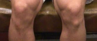
At the beginning of the disease, excessive movements that outwardly look like violent movements are random, at first glance, in nature. During initial diagnosis, it is important to correctly assess such symptoms. This will allow you to quickly make a correct diagnosis and prescribe adequate supportive therapy.
Outwardly, a person at the initial stage of development gives the impression of a person who, at the everyday level, simply cannot sit in one place. Then more and more muscle zones begin to be involved.
The gait becomes dancing. Further, the muscles of the pharynx and larynx are involved in the process, which causes disturbances in speech functions. As a result, it becomes increasingly difficult for others to understand what the sick person is talking about. As symptoms develop, hyperactivity gradually turns into general weakness and inhibition of actions.
After this, an active destructive process begins in the tissues of the brain, the final stage of which becomes the disability of the sick person. At the same time, anger, aggression, antisocial and inappropriate behavior are often observed, due to which interaction with others becomes difficult or completely impossible.
Subsequently, the well-being of victims of Huntington's disease deteriorates to such an extent that they begin to require round-the-clock care:
- feeding liquid or crushed food containing sufficient nutrients;
- proper organization of daily routine and hygienic care for the patient;
- in later stages of the disease, feeding through a feeding tube will be required.
Most patients experience increased appetite, but they do not gain weight, but continue to lose it. This is due to the fact that the processes of absorption and assimilation of nutrients are disrupted in their bodies. Therefore, in all cases it is necessary to provide the patient with nutritious food, high in calories and complete from a dietary point of view.
If there are difficulties in providing constant and regular care, you should consult with a specialist about the possibility of placing a sick person in one of the institutions such as a hospice, boarding school or paid clinic, where the patient will receive everything necessary for a relatively normal existence for the rest of his life.
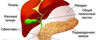
Pathogenesis
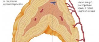
In this disease, there is a predominant lesion of the basal ganglia of the brain, which are of great importance in the control of movement and behavior. gamma-aminobutyric acid and substance P also decreases . These disorders occur due to gene mutations.
The Huntington's chorea gene is located on the fourth chromosome. The gene encodes the protein huntingtin (huntingtin), which is essential for cell development and survival. Normally, the gene contains a chain of cytosine-adenine-guanine (CAG) nucleotides, which is repeated. The number of such repeats in a gene is normally from 8 to 35, but in a mutagenic gene the number of repeats increases and this causes Huntington's disease. Because the CAG triplet encodes the amino acid glutamine , in this disease glutamine accumulates in excess and has damaging effects. The more CAG copies, the earlier the disease develops and the more severe the symptoms.
Models for studying Huntington's disease
Animal models of HD appeared more than 30 years ago. The first were models based on the introduction of neurotoxic substances into the striatum (for example, quinolinic acid, an NMDA receptor agonist), which caused neuronal death. Currently, most researchers are working on transgenic animal models, which include not only mice and rats, but also invertebrate animals - the fly Drosophila melanogaster and the worm Caenorhabditis elegans.
Mouse models of Huntington's disease differ from each other in the number of CAG repeats and the level of expression of the transgene - the artificially introduced huntingtin gene. Because The development of HD depends on these factors; different strains of mice differ from each other in the rate of development of pathologies. The most widely used models include the R6/2, R6/1, and YAC128 mouse strains, which were also used in our work. In mice of these lines, the symptoms of the disease are most pronounced and appear quite quickly. In addition, these animals develop cognitive and motor impairments with age and develop partial loss of neurons in the striatum and cortex.
Another way to model HD is to use cell culture. In the simplest case, cell cultures with stable transfection of the huntingtin gene are used. For example, these are PC12 cells containing an inducible transgene of the first exon of huntingtin or striatal neurons expressing huntingtin fragments of different lengths. In addition, primary cultures from neurons from transgenic mice or immortalized neurons can be used.
Classification
Extrapyramidal hyperkinesis is:
- Primary (for various neurodegenerative diseases, for example, Huntington's disease , neuroacanthocytosis , spinocerebellar ataxia ).
- Secondary (a symptom of autoimmune, vascular and metabolic diseases, heavy metal intoxication, encephalitis of various etiologies).
According to their prevalence, choreic hyperkinesis is divided into:
- Focal.
- One-sided.
- Generalized.
Huntington's chorea, what is it? This disease is associated with the destruction of neurons in the subcortical nuclei and cortex. The disease manifests itself as a combination of movement disorders, dementia and progressive cognitive impairment. Since chorea is not the only manifestation of the disease, the term "Huntington's disease" is more correct than the term "chorea". The disease appears at the age of 40-50 and affects both sexes equally. The main manifestations of the disease are steadily progressing.
Photos of symptoms
Already at an early stage of Huntington's disease, in addition to motor disorders, a pronounced speech disorder (slow arrhythmic speech) appears; a swallowing disorder appears in the later stages and becomes the cause of aspiration , asphyxia and pneumonia . Changes in the mental sphere are represented by impaired memory, attention, phobic disorders and depression with suicidal attempts.
Among the secondary forms, minor chorea (synonymous with Sydenham's chorea), which is a complication of rheumatism, . This is a generally recognized variant of rheumatic lesions of the nervous system, which is why rheumatic chorea is also called neurorheumatism . The first descriptions of an epidemic of rheumatic chorea were made in 1418, and a complete description of the disease was given by Sydenham 200 years later. In 1831, Bright established a connection between minor chorea and rheumatic fever and described severe forms of pseudoparalytic minor chorea.
In recent years, thanks to antirheumatic treatment, Sydenham's chorea is very rare, and rheumatism itself is considered by rheumatologists as a disease that is losing its relevance. In the early 80s, neurorheumatism (St. Vitus' dance) was diagnosed in 36% of children with rheumatism, and currently in 16% of patients. In this case, hemichorea (hyperkinesis of one side) and muscle hypotonia (moderate and severe), impaired coordination, handwriting and speech, increased tendon reflexes and emotional lability predominate.
Why we decided to study the parameters of synaptic transmission in Huntington's disease
Synaptic transmission is the transmission of signals between neurons using synaptic contact. When one neuron is excited, its synaptic ending releases a mediator into the synaptic cleft - a chemical substance that exerts its excitatory or inhibitory effect on the synaptic ending of the second neuron (Fig. 1). Thus, synapses connect neurons with each other, ensuring the normal functioning of neural networks and the entire nervous system. If any of the brain systems stops functioning, the reason may lie either in a disruption in the functioning of individual neurons, or in a disruption in the communication between them, i.e. disruption of synaptic transmission.
Figure 1. Schematic representation of the synapse structure.
"Wikipedia"
Huntingon's disease affects a specific area of the brain called the striatum. The striatum is part of an important neural pathway, the extrapyramidal system, which is involved in controlling movement and maintaining muscle tone. The death of striatal neurons in Huntington's disease leads to the destruction of the extrapyramidal system, which is associated with loss of control over movements in the sick person. But when the first pathological symptoms appear (tremor, loss of coordination), the human brain is not yet damaged: neurons begin to die only several years after the onset of the disease. Those. the disease begins when something changes in the functioning of the neurons themselves or in synaptic transmission, and these disturbances subsequently lead to the death of neurons and irreversible consequences.
The research results accumulated in recent years make many scientists inclined to believe that it is the disruption of the normal functioning of the neuronal communication system, synapses and synaptic transmission that leads to early disturbances in the functioning of the extrapyramidal system. It turned out that neurons with mutations in the gene encoding the huntingtin protein exhibit a number of pathological changes that disrupt synaptic transmission. In such mutant cells, the formation and renewal of the supply of vesicles (vesicles with a mediator) is disrupted, the intracellular concentration of calcium, which is necessary for the normal release of the mediator into the synaptic cleft, changes, the amount of a number of proteins necessary for the functioning of the synapse is reduced, etc. [7]. All this leads to a reduced release of the transmitter into the synaptic cleft, and if there is not enough transmitter, then the neurons begin to “hear” each other worse, and the commands sent by the cerebral cortex will not be carried out to the fullest extent.
In 2013, the Nobel Prize in Physiology or Medicine was awarded to works that made clear the details of vesicular transport - the process of formation and transportation of membrane vesicles (vesicles) between cells: “Nobel Prize in Physiology or Medicine (2013): vesicular transport” [8]. - Ed.
The study of impaired synaptic transmission in HD was the topic of our study. Could it be that the malfunctioning of striatal neurons in the early stages of HD is caused by the fact that they do not “hear” the commands of cortical neurons? Can weakening of synaptic connections lead to irreversible changes in striatal neurons and lead to their death? What we learned while searching for answers to these questions is described below.
Causes of Huntington's chorea
The cause of this disease is a genetic defect in the fourth chromosome. The genetic causes of Huntington's chorea are changes in the 4th chromosome, in which the number of repeats of one of the DNA fragments that encodes the protein huntingtin . The greater the number of repetitions, the earlier the disease develops. These genetic causes and the associated disruption of protein formation due to polyglutamine repeats determine the death of neurons in the striatum and caudate nucleus.
If we talk about secondary forms of chorea , their causes in childhood can be rheumatism , systemic lupus erythematosus , vascular damage ( vascular chorea ), and antiphospholipid syndrome . In the elderly, chorea can be caused by liver disease, stroke , and polycythemia . Chorea is the most common post-stroke hyperkinesis and is observed in 1.3% of patients with acute cerebrovascular accident. Choreic hyperkinesis appears acutely in the first days of a stroke . It is more often represented by hemichorea, and in the case of bilateral vascular lesions it is generalized. In most patients, hyperkinesis is combined with muscle weakness on the same side.
The cause of vascular chorea is ischemic or hemorrhagic damage to the thalamus , subthalamic nucleus and lentiform nuclei . Vascular chorea can also be caused by damage to the parietal, frontal or temporal lobes.
How to properly treat the disease
Unfortunately, at present there are no ways to completely get rid of the disease: doctors and scientists around the world do not yet have the ability to reprogram the human genome and force it to destroy the altered proteins.
A wide range of different supportive measures are now offered to patients with Huntington's chorea to improve their quality of life and overall well-being. Treatment is carried out both in a hospital setting and at home. Most patients require specialized care and nursing at the end of their lives, forcing them to seek special medical care.
As the disease progresses, the patient loses more and more independence, which leads to complete disability.
Principles of treatment of Huntington's disease:
- reduction in the frequency and intensity of attacks of hyperkinesis;
- relieving muscle spasm;
- elimination of convulsive syndrome;
- normalization of muscle condition with the help of therapeutic physical training;
- taking antidepressants to improve mood;
- using physical therapy to relax muscles;
- visiting a psychotherapist and psychiatrist.
Drug treatment
All medications intended to treat the disease must be prescribed by a doctor. After contacting a neurologist, the patient and his relatives receive a specific list of medications, which will subsequently be expanded.
It is not possible to cope with the disease on your own: only the doctor has the right to prescribe the necessary dosages that will help avoid deterioration of the patient’s well-being.
Table: drugs used to treat Huntington's disease
| Name of drug group | Examples of drugs | Effect of use |
| Antispasmodics |
|
|
| Antipsychotic medications |
|
|
| Sedatives |
|
|
| Antidepressants |
|
|
| Antiepileptic drugs |
|
|
Photo gallery: medications used in the treatment of chorea
No-spa relieves muscle spasms
Corvalol is a sedative that prevents the patient from becoming depressed
Phenazepam is prescribed to prevent seizures
Haloperidol can eliminate behavioral disorders and excessive physical activity
Fluoxetine reduces anxiety and helps lift the patient out of depression
Physiotherapy methods
Physiotherapy techniques are based on the influence of various physical factors on the human body. They help to relax the body and bring it into a calm state, as well as reduce the severity of hyperkinetic manifestations. A medical rehabilitation doctor draws up a program individually for each patient. The procedures are carried out over several months to ensure permanent results.
Table: Using Physical Therapy to Treat Chorea
| Name of physiotherapy technique | The essence of the procedures performed | Effects of treatment |
| Electrophoresis | Introduction of drugs into the body using direct current of varying strength | Fast and high-quality delivery of the drug to the human body |
| Darsonvalization | Application of low-frequency currents to stimulate specific muscle groups | Relaxation of spasming muscles |
| Magnetotherapy | Magnetic fields of varying degrees of strength affect the human body | Reducing the number and frequency of seizures |
| Laser therapy | Use of targeted laser radiation in the muscle area | Reducing the intensity of hyperkinesis |
Folk remedies
Traditional medicine recipes are widely used in neurology. Natural remedies are good sedatives, they are inexpensive and easy to use. But despite all the attractiveness of such methods, doctors do not recommend neglecting traditional treatment: without it, it will be impossible to achieve optimal results.
Here are a few recipes that can be used for Huntington's chorea:
- Take 50 grams of dried lemon balm and chamomile. Throw the resulting mixture into a saucepan with a liter of boiling water and cook for fifteen minutes. After cooling, drink 1 glass at night. Melissa and chamomile have a mild sedative effect. The course of treatment lasts at least six months with short (1-2 weeks) breaks.
- Purchase tinctures of ginseng and eleutherococcus at the pharmacy. Add ten drops of each tincture to your evening tea. These herbal preparations have the unique property of relaxing muscles and calming seizures. Treatment lasts three months, four times a week.
- Grate the valerian root on a fine grater. Brew the resulting mixture with a glass of boiling water and drink before bed. Regular use of valerian helps reduce the frequency of hyperkinesis, as well as reduce tremors. It is recommended to carry out this procedure three times a week for six months.
Photo gallery: components of folk recipes
It is recommended to collect lemon balm in August
Valerian root must be dried in the oven before use.
Eleutherococcus tincture can be purchased at the pharmacy
Homeopathic treatment
Homeopathy is a branch of alternative medicine that helps people fight various ailments using known chemical compounds and herbal preparations. In some cases, this approach is very effective: the number of seizures decreases, patients partially regain control of movements and can even voluntarily control facial expressions.
Any homeopathic medicine has special indications and contraindications for use. Be sure to consult your doctor before taking it: some medications may worsen your health.
The most famous homeopathic medicines that help with chorea:
- Agaricus muscarius is a remedy based on the red fly agaric, which is widely used in psychiatric practice to normalize the patient’s health.
- Alumina is a drug based on a compound of the metal of the same name, which allows you to stabilize the patient’s psyche and prevent the development of nervous breakdowns.
- Argentum nitricum based on silver nitrate is responsible for reducing the frequency of trembling attacks during illness.
Homeopathic medicines can be purchased at specialized pharmacies
- Hemlock is a homeopathic medicine made from the veh plant. It helps get rid of convulsive syndrome and relieves even the most severe muscle spasms.
Diet recommendations
The diet of patients with Huntington's chorea is also quite specific. Very hot, spicy, salty foods with a lot of chemical additives can negatively affect the patient's health. Particular attention should be paid to the type and shape of the food being prepared: it should not be too hard and hard, as it is difficult for patients to chew and swallow. It is recommended to prepare soups, purees and liquid porridges, and cut dense foods into small pieces.
The following foods should be excluded from the diet:
- canned food (meat, fish, fruit and vegetables);
- fatty meat (lamb, duck);
- fast food products (convenience foods, custard noodles and purees);
- fast food;
- carbonated drinks;
- store-bought juices;
- industrial sweets;
- alcohol of various strengths;
- crackers and chips;
- fish with a lot of bones.
Photo gallery: prohibited products
Fatty meat is poorly digestible
Carbonated drinks slow down your metabolism
Store-bought baked goods contain a large number of different additives
What to add to your diet:
- milk porridge (rice, buckwheat, oatmeal, semolina, rolled oats, millet, pearl barley);
- fresh vegetables, fruits and berries, chopped and pureed;
- milk and dairy products (cream, yoghurt, cottage cheese, cheese, kefir);
- lean meat and fish;
- seafood;
- nuts and dried fruits;
- soft sweet pastries;
- offal (liver, kidneys, lungs, ventricles);
- a large amount of water and homemade fruit drinks, jelly, compotes.
Photo gallery: products necessary for a patient with chorea
Dairy products replenish calcium reserves
Porridge is easily absorbed by the body and saturates it with essential vitamins and microelements
Vegetables and fruits are sources of vitamins
Methods of psychological rehabilitation
The disease often becomes a heavy blow not only for the patient himself, but also for his immediate family. They are forced to put up with constant mood swings, outbursts of aggression and irritability. Many patients have difficulty adapting to the changes that have occurred in their condition, which causes serious disturbances in behavior and perception.
For such cases, psychological and even psychiatric support and assistance are provided, aimed at stabilizing the patient’s moral health. The doctor helps to find out the causes of aggression, and also to put it in the right direction or even stop attacks of anger. The following methods are used:
- therapy with color and painting;
- sand painting;
- audiotherapy;
- directed work activity.
Patients suffering from Huntington's disease have the highest suicide rate. People fall into deep depression due to the loss of communication and self-care skills, which prompts them to commit suicide.
Each doctor seeks an individual approach to a patient with chorea and his relatives. First, it is necessary to explain to the family how to care for the victim, to warn about the possible risks and consequences of the disease. The patient is taught the basics of self-control, which helps him in the future cope with attacks of apathy and depression. Additionally, such patients are prescribed a visit to a speech therapist, who contributes to the partial restoration of lost speech function. Remember that you can go to a psychologist for both group and individual sessions. Communication in a team helps the patient to come to terms with his illness faster.
Symptoms of Huntington's chorea
Symptoms include various types of abnormal movements. They appear first in the distal parts of the limbs, and then become generalized, which disrupts the performance of normal movements. At first, patients experience sluggishness, clumsiness, poor coordination of movements, and handwriting deteriorates. Rapid and erratic movements in different muscle groups are associated with a decrease in the inhibitory influence of the brain on motor nerves.
Then there appears a bizarre gait (slow, tense, “dancing”), grimacing, inability to make a grasping movement and move the eyes without blinking or nodding the head. Patients cannot clench their hand into a fist. Over time, involuntary movements become dystonic and instability appears, which leads the patient to frequent falls. In severe cases, patients lose the ability to move independently and need a wheelchair. They also develop problems with swallowing and speech. Speech disorders are accompanied by involuntary muscle movements. When talking, the patient sobs, sniffles, smacks, grimaces, and the eyeballs move randomly. Later, speech becomes completely slurred.
In 60% of cases, psychopathic disorders appear before the onset of movement disorders.
Huntington's disease: symptoms and signs, photos
Emotional disturbances and increased excitability are manifested by the patient having unmotivated attacks of panic , rage , and anxiety . Hypersexuality may also develop . Sleep disturbances are noted.
Dementia develops in the later stages of the disease. Sometimes the development of dementia is stopped, and it does not always become total. Therefore, we can say that dementia in Huntington's chorea is relatively benign. Some patients can perform their usual work for a long time and remain outside the psychiatric hospital. Intellectual disorders manifest themselves in a decrease in the ability to think, weakening of attention and a lack of criticism of one’s behavior. If we consider other signs, we should note the high susceptibility of patients to depressive disorders and suicidal . Delusions and obsessive states may appear. Epileptic seizures may occur - most often this occurs in patients with the onset of the disease at a young age. Huntington's disease can be combined with diabetes mellitus . Therefore, such patients often exhibit symptoms of diabetes: increased thirst, increased amount of urine, dry mouth.
Depending on the predominance of a particular symptom in the clinic, several clinical forms of the disease are distinguished:
- Hyperkinetic . Involuntary muscle movement predominates, increasing with excitement. When walking, patients wave their arms, dance, sway, grimace, and make strange sounds when talking. Hyperkinesis disappears in sleep. Over time, they increase and patients are not able to care for themselves.
- Mental form . In patients, apathy, decreased criticism, memory impairment, agitation, delusional ideas, and hallucinations (auditory and visual) come to the fore. Dementia gradually develops.
- Akinetic-rigid form . This form is manifested by muscle rigidity and contractures. There are attacks of epilepsy , athetosis (movements in the distal parts of the arms and legs), oculomotor disorders, ataxia and dystonia . Mental development disorders are also noted.
Symptoms of minor chorea in children include “fancy movements”, hyperkinesis in various parts of the body, which impairs the performance of active movements and coordination of movements. Hyperkinesis in the facial muscles (grimacing, twitching) is characteristic. Children look restless and restless. The severity of hyperkinesis can vary - from slight twitching (if mild) to a “motor storm”.
Hyperkinesis of the oropharyngeal muscles is manifested by impaired swallowing and speech. Children choke when eating, and their speech becomes slurred and abrupt. 30% of children have unilateral hyperkinesis. In this case, muscle hypotonia is noted and tendon reflexes are inhibited. Emotional lability is also expressed: irritability and tearfulness are replaced by giggling and foolishness. Minor chorea develops in children 2-5 months after streptococcal infection and an infectious history plays a central role in diagnosis.
An important place is occupied by systemic manifestations of rheumatism : shortness of breath, palpitations at rest, enlarged liver , joint pain, the presence of ring-shaped erythema on the skin. Heart lesions ( endo- and myocarditis ) are detected on ECHO cardiography: enlargement of the heart chambers, changes in the heart valves, dysfunction of the valve leaflets. That is, neurological symptoms and signs of endomyocarditis .
Treatment
Huntington's disease has no specific treatment. The use of medications is aimed at eliminating its symptoms. As a rule, patients are observed by a neurologist, psychologist and therapist. Doctors prescribe complex treatment, including several groups of drugs.
To reduce the severity of muscle tension (rigidity), myoclonic hyperkensia (twitching) and hypokinesia (involuntary movements), antiparkinsonian drugs are used:
- levodol;
- valproic acid;
- pergolide;
- bromocriptine.
Treatment of depression and elimination of sleep disorders is carried out with the help of selective substances that suppress (inhibit) the reuptake of the neurotransmitter serotononin. Among them: Prozac, Zoloft and citalopram. Mirtazapine is also used.
For anxiety, aggression, psychosis and cognitive impairment, atypical antipsychotics are prescribed - amisulpride, clozapine and risperidone.
There is a constant search for effective pharmacological agents that can comprehensively influence the manifestations of Huntington's disease. In 2008, the drug tetrabenazine was approved for market in the United States. It reduces the degree of physical impairment, but has side effects: it can cause depression, drowsiness, dizziness, anxiety, akatasia (the inability to remain at rest for a long time).
Cognitive disorders require correction with the help of psychotherapy and training. A person with Huntington's disease needs emotional support.
In the terminal stage of the disease, it is important to provide proper care for the patient, since he completely loses the ability to make independent decisions and actions. If necessary, hospitalization is carried out.
Currently, studies are being carried out on the effectiveness and safety of drugs aimed at the preventive treatment of Huntington's disease: glutamate receptor antagonists and substances that activate the functions of the mitochondrial electrical transport chain. The action of these drugs is to protect neurons from the toxic effects of the mutant huntingtin protein. It is assumed that their use will produce positive results: delaying the onset of the disease and reducing the rate of its progression.
Tests and diagnostics
- Huntington's syndrome, as a hereditary neurodegenerative disease, requires medical and genetic examinations. Genetic testing is carried out - direct DNA diagnostics by fragment analysis. This makes it possible to identify carriage of a pathological gene in future parents and give an accurate prognosis regarding the health of the offspring.
- MRI and CT scans reveal atrophy of the caudate nucleus and atrophy of the cerebral cortex (more in the frontal lobe), but with a genetically confirmed diagnosis, these examinations have no diagnostic value.
When examining patients with rheumatic chorea, the following is revealed:
- Leukocytosis.
- Neutrophilia.
- Acceleration of ESR.
- Increased C-reactive protein and seromucoid .
- Increased titers of antistreptolysin O (from 1:500 to 1:1000).
- ECHO-CG shows an increase in heart size, changes in valves.
Types and forms of pathology
Huntington's disease is a pathology whose incidence does not exceed 1 case per 10,000 people in the general population . This largely depends on the race and area where people live.
There is no special division into types. The disease is characterized only by stages of development and the severity of existing dysfunctions of individual parts of the human body.
Chorea in children
Minor chorea in children, which is a complication of rheumatism , has rarely occurred in recent years, as has rheumatism itself. This is due to the effective treatment of sore throats and rheumatism through the timely and rational use of antibiotics.
In the clinical picture of rheumatism, there is a “latent” period between tonsillitis or pharyngitis and the appearance of fever, intoxication, weakness, and the development of carditis , arthritis and chorea . Numerous symptoms indicate systemic inflammation of the connective tissue localized in the heart, joints and brain. There is a rule: the presence of carditis or chorea can be considered as probable rheumatism and treatment can begin.
Symptoms of minor chorea in children include the appearance of chaotic, involuntary rapid movements that occur when dressing, playing, eating and walking. The movements involve the proximal parts of the arms and legs and intensify with emotional stress. Hyperkinesis begins smoothly compared to Huntington's chorea, but also affects the muscles of the pharynx, face, diaphragm, and trunk. Lesser chorea has a favorable course. The duration of the disease is 3-6 weeks. When the course is not severe, movement disorders completely disappear. In severe cases, a long course of up to several months and the presence of residual effects in the form of tics are possible.
In the treatment of rheumatism, attention is paid to eliminating the pathogen by intramuscular administration of penicillin . For allergies to penicillin, macrolides ( Erythromycin , Azithromycin ) are used. Non-steroidal anti-inflammatory drugs ( Nise , Ibuprofen , Airtal , Meloxicam ) are prescribed for three weeks. For carditis, glucocorticoids are prescribed for up to 15 days, and then the dose is reduced by 20% every week. Glucocorticoids are ineffective in the treatment of chorea. Children are prescribed Haloperidol (0.5-1.0 mg per day, increasing the dose by 0.5 mg every three days). Sodium valproate is effective ; in resistant cases, plasmapheresis, Etaperazine and Reserpine .
Antiphospholipid syndrome also occurs in children . This is a systemic autoimmune disease manifested by high titers of antibodies to phospholipids, arterial or venous thrombosis . A high titer of antibodies to phospholipids causes pathology of the central and peripheral nervous system: convulsions , epileptic seizures , transient ischemic attacks , partial chorea , hearing loss , amnesia and psychosis .
Optogenetics
Optogenetics is a method that combines the approaches of genetics and optics for fine control of the electrical activity of electrically excitable cells (neurons and muscle fibers) [9]. To do this, genes of special light-sensitive proteins—microbial opsins, which are ion channels or pumps—are introduced into the cells under study (Fig. 7). The first work showing the possibility of controlling the electrical activity of neurons using opsin was published in 2005. Over the next few years, a number of experimental works appeared that made it possible to refine this technique and prove its applicability under various experimental conditions.
In recent years, many different opsins have been discovered, of which halorhodopsins and channelrhodopsins have found the most application in optogenetics. When the opsin gene is delivered using genetic engineering methods into a neuron, light-sensitive channels appear on the plasma membrane, and the cell itself becomes light-sensitive. When exposed to blue light, the channelrhodopsin pore opens (maximum absorption - 470 nm), which causes the movement of positively charged ions into the cell, ensuring depolarization of the neuron membrane and the generation of action potentials. When exposed to yellow light, halorhodopsin is activated (maximum absorption - 580 nm), the neuron membrane is hyperpolarized, causing inhibition of the neuron. The high temporal resolution of optogenetics allows for very fine regulation of synaptic events and is therefore an important tool for studying interneuronal connections.
The combined use of optogenetics and classical electrophysiology techniques allows us to benefit from the positive qualities of each of these approaches. The precision of electrophysiological recording is combined with the ability to use light stimuli of varying durations and intensities to help scientists study the workings of neural connections in detail.
The electrical activity of neurons is expressed in sharp voltage surges on the cell membrane and the resulting appearance of an electric current. These sudden jumps are called spikes or action potentials and last a few milliseconds (Figure 8). We found that the longer a neuron is illuminated with blue light, the more spikes it creates during this time. If the illuminated neuron is connected by a synaptic contact with another neuron, then response activity can be recorded on the second neuron, also in the form of individual spikes.
Figure 8. An example of a recording obtained from electrophysiological recording of neuron activity. Spikes (action potentials) are reflected in the recording as vertical lines showing sharp jumps in membrane current.
In our experiments, we used a pair of young (14 days) cortical and striatal neurons connected by synaptic contact. The cortical neuron was activated with blue light, and the response activity was recorded on the striatal neuron. It turned out that for a response to occur on a striatal neuron, a certain duration of illumination (activation threshold) is required. If the duration of illumination was below the threshold value, then a spike on a striatal neuron did not occur in response to every flash of light. And most importantly, the activation threshold for neurons from the brains of healthy mice was different from YAC128 mice (with a mutation in the huntingtin gene). This difference was most clearly visible at 50% activation of the striatal neuron, i.e. the duration of illumination at which a spike occurs in response to every second flash of light. The threshold for 50% activation of a striatal neuron in response to irradiation of a cortical neuron for the YAC 128 cell culture was approximately two times (more precisely 2.3 ± 0.8) higher compared to the positive control [10].
It turns out that a mutation in the huntingtin protein leads to the fact that synaptic transmission deteriorates even in young neurons, and the disturbances occur at the functional level (without morphological changes). Could it be that it is these functional disorders that subsequently lead to the appearance of morphological changes, such as the disappearance of spines in old striatal neurons?
To answer this question, we again turned to optogenetics for help, but now with its help we suppressed the activity of neurons with light (Fig. 9a). If our assumption is correct, then the temporary absence of the activating influence of the cortex should lead to the disappearance of spines on striatal neurons in the YAC128 culture. Indeed, after the experiment, the number of spines in the positive control remained unchanged, but in the YAC128 culture it decreased significantly (Fig. 9b, c). It turns out that neurons modeling HD are especially sensitive to weakening the activating influence of cortical neurons , therefore, long-term weakening of the synaptic connection between these neurons leads to a decrease in the number of spines on the 20th day of cultivation [10].
Figure 9. Influencing neurons using optogenetics. a — suppression of cortical neuron activity using optogenetics. When a neuron is illuminated with yellow light (orange stripe), it becomes inactive—there are almost no spikes in the electrophysiological recording during this period of time. b — influence of long-term optogenetic inhibition on the morphology of neurons. Micrographs show a decrease in the number of spines on the dendrite of the YAC128 neuron. c — change in the density of dendritic spines after optogenetic inhibition. In the wild-type culture, the number of spines does not change, but in the YAC128 culture, spine density is significantly reduced.
In summary, we found that in our HD model there is a disruption of synaptic transmission that develops in two stages. At an early stage (young neurons, on day 14 of neuron culture), a functional weakening of the synaptic connection occurs, which at a later stage (old neurons, on day 20 of culture) leads to morphological disturbances of synaptic contacts.
Diet
In the initial stages of the disease, when swallowing is not impaired, the diet of patients does not differ from the diet of healthy individuals. In patients with advanced manifestations, swallowing is impaired and the ability to eat normally is significantly limited. Such patients quickly lose weight and have high energy consumption. In this connection, an important aspect is the regulation of the nutrition of patients.
It is recommended to consume higher-calorie foods in pureed and mushy form to minimize the difficult chewing process. It is recommended to increase the total caloric content of the diet by 500 kcal. In addition, vitamin therapy, the use of mineral supplements and biologically active substances are indicated. In some countries, PUFAs (omega 3 fatty acids) are widely used in the form of food supplements for neurological pathologies, including Huntington's disease.
Research Findings: Changes in Synaptic Transmission in Huntington's Disease
Synaptic transmission can be studied in various ways. For example, this can be done by penetrating the neural circuit using electrophysiological methods. A neuron expresses its activity using an electrical current that can be measured. If an experimenter takes a chain of two neurons and, after activating one neuron, records the electrical activity of the second, he can find out how well the signal travels. Another way to study the functioning of synaptic transmission is to study the morphology of the neuron. The fact is that many neurons (including neurons of the cortex and striatum) have special membrane outgrowths - spines, which they need specifically for the formation of synapses (Fig. 2). The more actively a neuron “communicates” with its neighbors, the more spines there are on its surface. Taking these two approaches, we decided to investigate how synaptic transmission works in HD.
Figure 2. Dendritic spines on the surface of a striatal neuron. Spines are small projections on the surface of neuronal processes; in the enlarged image they are marked with arrows.
photo of the author of the article
Figure 3. Neuronal culture from cortical and striatal neurons. Using specific antibodies, cortical neurons are colored red, and striatal neurons are colored yellow-green.
photo of the author of the article
As a model for studying Huntington's disease, a cell culture from neurons of the cortex and striatum was used. To prepare the culture, immature neurons from the studied areas of the mouse brain are planted in Petri dishes, where they form full-fledged neuronal processes and neural chains (Fig. 3). Wild-type mice (without mutations) and YAC128 mice, which carry a mutation in the huntingtin protein gene and are a recognized model of HD, were used. On days 14–15 after neurons are planted in a Petri dish, they reach a mature state corresponding to the state of neurons in the adult brain, and on days 19–20 neurons are considered “old”: they exhibit a number of cellular processes characteristic of the brain of elderly people. In addition, with age, mutant huntingtin protein and its aggregates accumulate in neurons of YAC128 mice, so studying neuronal culture at these two stages provides insight into what is happening in the brain of a patient with HD at the early and late stages of the disease.
Figure 4. Different types of spines on the surface of a dendrite - micrograph and schematic representation.
To begin with, we examined the morphological differences between the two lineages, i.e. compared their appearance. Normal striatal neurons are characterized by the presence of a large number of spines, which is why they are called medium spiny neurons (MSNs). It is the spines that form the majority of synaptic contacts between neurons of the striatum and cortex, and the presence of a certain number of them is important for normal synaptic transmission. The “quality” of spines is also important: in modern neurobiology they are divided into three groups according to size and shape (Fig. 4): mushroom-shaped, thin and stump-shaped. At the same time, different types of spines perform different functions: it is believed that only mushroom-shaped spines form active synapses, while thin and stump spines do not form contacts with other neurons. Thus, for the normal functioning of the neural chain and the effective transmission of information along it, the presence of a certain number of mushroom-shaped spines is necessary.
In order to find out how many mushroom-shaped spines there should be on the SCN normally, a culture of neurons from the brain of wild-type mice was used as a control at all stages of the morphological analysis. It turned out that the number of spines on the SSN of the striatum on day 14 of culture (young neurons) is the same in YAC128 and wild-type cultures, but on day 20 (in “old” neurons) significant changes are observed (Fig. 5). In an “aged” YAC128 culture, the total number of spines decreases, and the relative number of mushroom-shaped spines that form active synapses decreases by half [10]. It turns out that morphological changes in neurons, indicating a violation of synaptic transmission, develop only in “old age” (at a late stage of the disease). This means that in the early stages the root of the problem must lie in another area.
Figure 5. Morphological analysis of striatal neurons. a — neuron spines on days 14 and 20 of culture. Micrographs show areas of neuron dendrites. The relative number of spines of different types is marked on the pie chart: green - mushroom-shaped spines, red - thin spines, black - thin spines. On the 20th day of cultivation, the proportion of mushroom-shaped spines on the surface of YAC128 neurons decreases and the proportion of stump spines increases. b — Dendritic spine density (average number of spines per 10-μm-long dendrite) on wild-type and YAC28 neurons.
Therefore, we compared the electrophysiological characteristics of the two lines. This is possible thanks to the electrical activity of neurons, which can be recorded using a special method called patch clamp. To do this, a thin glass pipette with an electrode inside is applied to the surface of the neuron. When a neuron is activated, the voltage on its cell membrane changes, and this change is recorded by an electrode. If you take two neurons connected by a synaptic contact and, exciting one, record the response electrical activation on the second, you can measure the efficiency of signal transmission through the synapse (Fig. 6). If response activation does not always occur, then synaptic transmission is likely weakened. This method can detect synapse dysfunction before changes in neuron morphology.
Figure 6. Experimental design using combined optogenetics and electrophysiological recording. The cortical neuron is activated when illuminated with blue light, and the response activity is recorded on the striatal neuron in contact with it using a glass pipette with an electrode.
To activate a neuron, you can use several methods. A chemical can be added to the surrounding fluid to open ion channels, causing the neuron to become electrically excited. You can stimulate a neuron with an electrical current. But for our experiments we chose a more subtle tool for influencing neurons, namely optogenetics. This approach is based on the introduction of special light-sensitive proteins - opsins - into neurons, as a result of which the neurons themselves become sensitive to light (Fig. 7). As a result, by illuminating neurons with light of a specific wavelength, their activity can be changed. Illumination with blue light excites the neuron, and with yellow light it causes inhibition (i.e., it suppresses the activity of the neuron).
Optogenetic technologies these days even promise to restore vision to people with degenerative retinal lesions, imparting light sensitivity not to destroyed photoreceptors, but to ganglion cells: “Optogenetics + holography = insight?” [eleven]. - Ed.
Figure 7. Opsins are light-sensitive proteins that enable the movement of ions across the cell membrane and changes in neuronal activity. Channelrhodopsin (ChR2) causes membrane depolarization and neuron activation, halorhodopsin (NpHR) causes membrane hyperpolarization and neuron inhibition.
Prevention
There is no prevention for Huntington's disease because it is a genetically determined disease. It is impossible to prevent the disease in mutation carriers. People of childbearing age who have relatives with this disease must undergo genetic testing. If a carrier of a mutation is detected, it is recommended to avoid childbearing. As for rheumatic chorea, the following measures are the prevention of rheumatism and, accordingly, its complications in the form of hyperkinesis:
- hardening;
- good nutrition;
- rational physical education;
- walks in the open air;
- carrying out sanitary and hygienic measures that reduce the risk of streptococcal infection of children in the team;
- timely and effective treatment of sore throat and pharyngitis ;
- year-round bicillin prophylaxis for rheumatism .
The standard antibiotic treatment regimen for sore throat should be 10 days. The optimal penicillin drug is Flemoclav Solutab (amoxicillin/clavulanate). In case of poor intolerance to β-lactam antibiotics, macrolides ( Azithromycin , Clarithromycin , Josamycin ) are prescribed. Secondary prevention of repeated attacks of rheumatism involves year-round administration of long-acting penicillins ( Extencillin , Retarpen , Bicillin 5 and Bicillin 1 ).
The duration of secondary prevention can be 5 years to lifelong use (for patients with heart disease). An effective dosage form is Extensillin , which has advantages over Bicillin-5 . Of the domestic drugs, Bicillin-1 (injections once a week).
Causes of the disease and risk factors
Rheumatic chorea is caused by group A streptococcus. That is why sore throat is one of the main causes, after which all those who have recovered automatically fall into the high-risk group. Another likely cause is infectious diseases of the upper respiratory tract. But not everyone can develop a serious complication due to a sore throat.
The following conditions are considered the main risk factors:
- insufficiently strong immunity;
- cerebral palsy;
- poor blood circulation in the brain;
- carious lesions of teeth;
- hormonal imbalance;
- other foci of rheumatism;
- some drugs.
Against the background of these factors, the development of chorea is likely due to a violation of the functional abilities of ganglia in the brain structures and dentate nuclei of the cerebellum.
Consequences and complications
After 15-20 years, patients develop complications:
- Dysphagia , in severe cases of which the possibility of percutaneous endoscopic gastrostomy (installation of a feeding system in the stomach) is considered.
- Loss of body weight up to cachexia , which is associated not only with impaired swallowing, but also with cognitive impairment, when the patient loses the skills of eating or forgets to take it.
- Aspiration pneumonia due to dysphagia.
- Heart failure.
Provoking factors
Factors that provoke the early onset of Huntington's chorea include:
- alcohol abuse;
- smoking;
- drug use;
- unhealthy diet (excess fried, salty foods);
- sedentary lifestyle;
- excess body weight;
- work in hazardous and harmful production (contact with chemicals, radiological contamination, emissions);
- living in environmentally unfavorable areas.
List of sources
- Shtok V.N., Ivanova-Smolenskaya I.A., Levin O.S. Extrapyramidal disorders: Guide to diagnosis and treatment. - M.: Medpress inform, 2002. - 700 p.
- Shtok V.N., Levin O.S. Drug-induced extrapyramidal disorders //In the world of drugs. - 2000. - No. 2. - P. 3-7.
- Seliverstov Yu.A. and others // Nervous diseases. 2014. No. 3. P. 24.
- Tyurin N.A., Artamonova V.A., Alexandrova K.A. and others. Features of the modern course of minor chorea in children. Pediatrics. 1987; 2:20–23.
- Pediatric rheumatology (Guide for doctors) / Ed. Baranova A.A., Bazhenova L.K., 2002; 49.