Wherever bones, tendons, or ligaments move and rub against each other, especially in the joint area, the points of contact are cushioned by small fluid-filled sacs called bursas. The bursae are lined with special cells called synovial cells that produce a fluid rich in collagen and protein. By reducing friction, each of these bags (there are about 150 of them in the body) helps the joints work, smoothly providing the necessary range of motion. The bursa allows for multi-vector movements in joints such as the shoulder, elbow, knee, femur, and ankle. Inflammation and swelling of the bursa is called bursitis .
Bursitis of the knee joint
Symptoms of knee bursitis vary depending on the specific location of the inflammation, as well as the nature of the exudate formed.
Bursitis of the knee joint
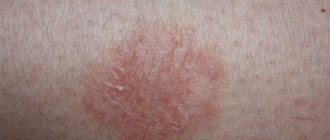
If the form is serous, slight swelling and tissue hyperemia will be observed. On palpation, the formation feels elastic. During examination, the patient notes moderate pain. The main cause of complaints is stiffness of movement, malaise, and decreased performance.
If it is purulent bursitis, then pain and increased temperature at the site of development will be high. When palpated, the formation will be dense and hot. In advanced cases, palpation may also reveal deposited salts.
Treatment consists primarily of emptying the cavity of the synodal bursa from excess fluid. If an infection is suspected, antibiotics are injected into the cavity. Anti-inflammatory drugs, compresses, and ointments are prescribed that relieve swelling, help normalize blood flow in the tissues and reduce inflammation and pain.
Bursitis of the knee joint is divided into several types:
- Suprapatellar bursitis is localized at the junction of the knee cartilages.
- Anserine bursitis - inflammation of the knee joint itself. It is characterized by severe pain and difficulty in flexing and extending the joint.
- Infrapatellar bursitis of the knee joint. Appears mainly after injury to the kneecap. There are as many as two infrapattellar bursae located in the human knee. The first is subcutaneous, the second is located deeper, between the muscle and the bone, opposite the subcutaneous bursa.
- Foot anserine bursitis is a deep type of bursitis. The knee joint is not affected. Localization - the inner surface of the knee, the place of attachment of the internal collateral ligament, semitendinosus and semimembranous muscles. The so-called tendobursitis of the pes anserine tendons. Like when walking. Likewise, when standing for a long time, severe pain is felt when bending on the inside of the joint.
A meniscus cyst can be mistaken for a manifestation of bursitis. It also develops in connection with trauma and increased stress. The signs that a cyst manifests itself are tumor-like compactions on the outer surface of the knee joint.
If treatment is not timely, the cyst can grow and deform adjacent tissues. At the initial stages, the cyst is treated conservatively with injections, but if the results are not satisfactory, knee surgery is prescribed to remove the formation and the tissues modified under its influence.
Pathological anatomy
Pathoanatomical changes in acute B. are expressed by signs of acute inflammation of the walls of the synovial bursa.
The initial stages of acute B. are characterized by serous impregnation of tissues and accumulation of serous exudate in the cavity of the bursa (acute serous B.). In the presence of microbial flora, serous inflammation quickly turns into purulent inflammation (purulent B.). The spread of the purulent process to surrounding tissues can occur as a phlegmonous inflammation with necrosis of the bursa wall and the formation of subcutaneous and intermuscular phlegmon. In advanced cases, fistulas that do not heal for a long time are formed. The breakthrough of pus into the joint cavity leads to the development of purulent arthritis.
In acute traumatic bursitis, hemorrhagic fluid accumulates in the stretched synovial bursae and their pockets. With reverse development, fibrin is organized and the vessels of the synovial membrane are obliterated. Persistent changes develop in the wall of the bursa, the edges thicken, the surface of the synovial membrane is covered with growths of connective tissue (proliferating B.), dividing the cavity of the bursa into additional pockets.
A microscopic examination of the wall of the bursa in places where connective tissue grows reveals its hyaline degeneration; Cell proliferation is observed around the vessels, and in places there is inflammatory infiltration of the vascular walls.
The deposition of salts in the wall and cavity of the bag leads to the development of the so-called. calcareous B. (bursitis calcarea), and the organized fibrin forms the so-called. villous (“rice”) bodies.
When acute inflammation subsides and during the subacute course of B., encapsulated areas of necrotic tissue or exudate remain in the wall and pockets of the bursae, which, with repeated injury and infection, serve as fertile ground for the development of relapse of inflammation (recurrent B.).
In tuberculous B., the walls of the bursa are evenly thickened, and tuberculous tubercles are found in it.
Elbow bursitis
The elbow joint is quite often susceptible to inflammatory processes, as it is actively involved in sports, outdoor games and physical activity in everyday activities. Articular bursae can become inflamed due to inadequate load, or if an injury occurs.
Symptoms indicating elbow bursitis:
- The appearance of edema;
- the hand swells, but may remain painless;
- discomfort, pain in the joint area;
- redness of the skin in the elbow area;
- increased body temperature;
- swollen lymph nodes;
- inability to bend the arm at the elbow;
- formation of fistulas, phlegmon in case of suppuration in the joint.
Treatment consists primarily of immobilizing the limb - from a splint to a plaster cast. Acute bursitis is characterized by acute pain. Increased body temperature and intoxication. Purulent bursitis of the elbow joint develops under the influence of viruses and pathogenic bacteria. There is swelling. The patient complains of “bloating” in the elbow.
Broad-spectrum antibiotics are prescribed to suppress the infection. If there is exudate, pump out with a syringe and rinse the cavity. Non-steroidal anti-inflammatory drugs are indicated. The patient is prescribed immunostimulating drugs and vitamin complexes.
If no signs of relief are found, then surgery to drain the joint capsule is prescribed. The fluid is pumped out, the damaged bursa is removed, which will subsequently be restored during rehabilitation.
Diagnosis
The diagnosis is simple for inflammation of superficially located bursae and is based on the typical clinical signs described above. The diagnosis is facilitated by puncture of the bursa cavity, which makes it possible to determine from the resulting contents the nature of the inflammation (serous, purulent, purulent-hemorrhagic, etc.), to determine the nature of the microbial flora and its sensitivity to antibiotics.
To carry out the most effective treatment, it is important to exclude the specificity of the infection that caused the inflammation (gonococci, brucella, spirochetes, etc.), which is possible on the basis of a carefully collected anamnesis, bacteriological examination of the contents of the bags, and the results of specific serological reactions.
The main differential diagnostic feature to distinguish B. from arthritis is the preservation of movements in the joint.
Rice. 2. Achilles bursitis. An enlarged synovial bursa is detected, pushing the Achilles tendon posteriorly, and inflammatory infiltration of fatty tissue upward and anterior to it; marginal destruction of the bone bed of the synovial bursa of the posterior surface of the calcaneus. Rice. 3. Calcareous subacromial bursitis. An intense, shapeless shadow of calcareous deposits is visible in the subacromial bursa (above the head of the humerus). The structure of the areas of the greater tubercle of the humerus is sparse (local osteoporosis).
X-ray diagnostics
superficially located (subcutaneous) B. of any localization is relatively simple. Due to their availability for clinical recognition, it is usually only of a clarifying nature. X-ray diagnostics of deep blisters is of much greater practical importance. Among them, the radiologist most often encounters blemishes in the area of the knee joint and greater trochanter of the femur, Achilles bursitis and inflammation of the unstable subcalcaneal mucous bursa, and on the upper limb - with chronic, subacromial bacilli (Fig. 2 and 3).
The main method of radiological recognition of bursitis is radiography. Its diagnostic capabilities depend on the location of the affected mucous bursa, the nature and severity of changes in it, as well as the presence or absence of secondary changes in nearby bones. Deep B. is easier to recognize if the anatomical region where it occurs provides a well-differentiated picture of soft tissues on the radiograph (for example, the area of the knee joint, the calcaneus). In such cases, B.'s diagnosis is based on detecting the shadow of an enlarged synovial bursa and inflammatory infiltration around it (Fig. 2). On the contrary, if there is insufficient natural contrast in the image of soft tissues (the area of the shoulder joint, the greater trochanter of the femur), B., as a rule, is detected either by the deposition of calcareous salts in the wall or cavity of the bursa, or in the presence of various kinds of changes in the adjacent bones (Fig. 3 ).
In some cases, for the purpose of differential diagnosis of B. (hygroma) of the knee joint area, in addition to conventional radiography, it is necessary to carry out arthrography (see), which makes it possible to identify or reject the connection of the joint cavity with the identified pathological formation. To accurately recognize B. of certain localizations, artificial contrasting of the synovial bursae is sometimes resorted to by direct puncture (bursography). In this case, both positive and negative radiopaque agents are used.
Bursitis of the shoulder joint
Typical for people involved in sports or high physical stress on the shoulder joint. It occurs more often in men.
Symptoms of shoulder bursitis include redness and swelling. Pain may be felt with sudden movements of the hand. There is a feeling of limited movement.
Treatment is prescribed with anti-inflammatory and painkillers. A splint is often practiced to relieve stress on the shoulder.
There are several types of the disease:
- Subacromial bursitis of the shoulder joint. Occurs under the influence of frequent injuries and heavy load. The supraspinatus tendon is affected. When the shoulder is abducted at an angle of 60 to 120 degrees, increased pain is felt. In its advanced form it radiates to the shoulder and arm.
- Subdeltoid bursitis. The pain radiates to the shoulder and arm.
- Subcoracoid bursitis. The joint capsule under the coracoid process of the scapula is affected. Pain when moving the shoulder back.
- Subcoracoidal bursitis of the shoulder joint also occurs with excessive activity.
- Calcareous bursitis develops after dislocations and subluxations, most often in athletes.
- Calculous bursitis is characterized by such severe pain. That the only way to cope is by blocking the analgesic.
With all manifestations, the main signs are pain and redness of the shoulder. This alone should be the reason for a visit to the doctor, since only an examination will indicate a specific reason.
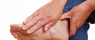
Ankle bursitis
The ankle joint suffers more from injury in men. Its damage in women is more associated with wearing uncomfortable and tight shoes.
Symptoms of bursitis on the ankle are swelling, redness, pain when moving, “bumps” and painful calluses may form.
Treatment is selected by the doctor based on the etiology of the disease and severity. Wearing splints, antibiotics, if necessary, cleaning the joint capsule or excision of the bursa. Medicines, injections, and physiotherapy are prescribed.
Bursitis of the foot often affects overweight people, since when walking, the joint is subject to regular excess load.
Bursitis of the metatarsal bone of the foot is felt by pain in the area of the sole and around the bone head. The foot may change in appearance and become swollen. Painful calluses form. Walking becomes very uncomfortable.
Diagnostics
The diagnosis of bursitis is made based on a combination of symptoms, clinical manifestations, medical history and instrumental diagnostic methods. The medical history allows us to determine the presence of concomitant somatic pathology.
Certain diagnostic procedures may be prescribed to rule out other causes of pain. They include the following diagnostic procedures:
- X-rays can visualize the presence of osteophytes or arthritis.
- Analysis of the punctate (microscopy), which was obtained as a result of puncture of the enlarged bursa, allows us to exclude gout and the presence of infection. Bursitis in the knee and elbow are most likely to become infected.
- Blood tests can help rule out rheumatologic diseases (such as rheumatoid arthritis) and metabolic diseases (diabetes mellitus).
- MRI may be prescribed if detailed visualization of the morphological picture is necessary.
Bursitis of the hip joint
The hip joints bear almost the majority of our body weight. Nearby there are a large number of attached muscles that are actively exposed to stress.
Symptoms are expressed in sharp, even burning pain when rotating the hip inward, squatting, or lying on the side. Felt on the front, outer surface of the thigh. At the moment of extension they intensify.
Treatment includes not only relief of inflammation and pain, but also physical therapy. Immobilization is indicated in acute forms or with the development of post-traumatic bursitis.
Trochanteric bursitis is the most common disease characteristic of women who are professionally involved in sports. A wide pelvis compared to a man's pelvis increases friction between the tendons. Long distance running increases the risk of developing the disease. An iliopectineal type of bursitis may also occur.
Basic methods of physiotherapy
Chronic bursitis is treated with physical therapy. It responds well to treatment with this method. Physiotherapy resolves tumors, relieves pain and relieves inflammation much faster when combined with medications. For medicinal purposes the following methods can be used:
- applications with paraffin;
- ultrasound;
- electrophoresis;
- inductothermy;
- microwave treatment;
- naphthalan baths;
- massage.
Each type of physiotherapy has indications and contraindications. Physiotherapy is prescribed after the patient is notified of possible side effects.
Ischiogluteal bursitis
Symptoms of inflammation of the ischial bursa (between the gluteus maximus muscle and the base of the pelvic bone):
- Pain when sitting and while walking, radiating to the pubic bone area;
- felt in the back of the thigh;
- when the hips are flexed passively, it is felt more strongly in the lying position.
- the patient cannot stand on tiptoes.
It is more common among cyclists and those who have been forced to sit on a hard surface for a long time.
Bursitis of the hand
The bursa of the wrist joint becomes inflamed.
Symptoms: severe swelling in the wrist; sharp pain extending to the shoulder; hand movements are limited; inflammation of the skin and local increase in temperature in the area of inflammation. Lymph nodes enlarge.
Treatment. The acute form is treated surgically by excising the bursa. First of all, the joint is fixed using a pressure bandage or splint. Nonsteroidal drugs and antibiotics are administered. They use Hydrocortisone or Kenalog - hormonal drugs that help relieve inflammation. Use ointments and gels.
Causes of bursitis
Inflammation of the bursa, or bursitis, develops as a consequence of bruise, injury, or infection of the body. The second factor is excess load on the joint. Bursitis of unknown etiology is also diagnosed when there are no apparent reasons for its occurrence.
Video on what causes bursitis:
Bursitis of the ankle and hip joint is rarely detected. Most often, the joints of the knees, elbows and shoulders suffer from the disease - since human activity mainly places the load on them.
Inflammation of the bursa can be caused by an injury with an abrasion, a wound, or even an uncomfortable shoe last. A very important factor is that exposure should be regular. In this case, the risk of developing bursitis becomes maximum. Mechanical damage also contributes - dislocations, subluxations and fractures. Autoimmune diseases. Disturbance of metabolic processes.
Etiology and pathogenesis
The cause of acute bursitis is most often trauma (bruise, abrasion, small wounds) and secondary infection of the synovial bursa with pyogenic microbes - mainly staphylococci and streptococci, much less often gonococci, pneumococci, tuberculosis and E. coli, Brucella, etc. Infection of the synovial bursa occurs through the lymphatic tract from purulent foci (with erysipelas, boils, carbuncles, osteomyelitis, bedsores), the hematogenous route of infection cannot be excluded. Chronic B. is often the result of prolonged constant mechanical irritation.
Treatment of bursitis
In case of inflammation of the joint capsule, treatment will be carried out comprehensively in order not only to remove the symptoms of the disease, but also to prevent relapse. For this purpose the following can be used:
- Injections - especially in acute forms, when it is necessary to remove inflammation and swelling, to overcome the infection that has arisen directly at the site.
- Pills. The doctor will prescribe anti-inflammatory drugs and antibiotics. The tablets, in parallel with the injections, affect the body as a whole, preventing the disease from affecting similar systems.
- Ointments and gels are used for topical use during the period of no exacerbation, when the manifestation of bursitis is moderate or mild.
- Physiotherapy is aimed at restoring the normal functioning of tissues, starting regenerative processes, and enhancing trophism when using other drugs.
Courses of treatment can vary from 10 days to a month, depending on the stage of development of the disease and aggravating symptoms.
Drug treatment of bursitis
When visiting a doctor about inflammation of the joint capsule, be prepared to take pills for bursitis. This is not the doctor's whim. The fact is that a painful process for the patient is associated with a whole complex of factors that should be eliminated:
- Inflammation - NSAIDs are prescribed;
- Painkillers are drugs that block pain receptors, designed to allow the patient to be at rest.
- Decongestants – the accumulation of fluid puts pressure on the receptors, which leads to additional discomfort. In addition, excess fluid in the tissues does not contribute to joint mobility.
- If the development of an infection is detected, then drugs will be prescribed to suppress pathogens.
Prescribed Dimexide for bursitis is used together with novocaine in the form of a warm compress. The drug can also be diluted with water. The purpose of the antiseptic is to rid the joint capsule of the accumulation of purulent exudate. It is sometimes used to promote better penetration of other drugs.
Dimexide is used for 10 days. Moreover, the maximum time for compresses is 40 minutes. In case of discomfort, the bandage is removed earlier to avoid burns.
Other compresses are also used for bursitis. A saline solution in the form of a compress acts on the principle of osmotic pressure and draws fluid from the joint. A compress of cabbage leaves and honey acts as an anti-inflammatory and decongestant.
In case of acute manifestations of bursitis, cover the sore spot with ice for 10 minutes, after which the joint is warmed with a warm compress based on a decoction of burdock, yarrow, and St. John's wort.
Treating bursitis at home
Often people do not want or for some reason cannot see a doctor. When pain occurs, treatment of bursitis with folk remedies involves the use of alcoholic and non-alcoholic infusions, compresses and ointments based on honey, propolis, other bee products, aloe, St. John's wort, yarrow and horse chestnut.
The action of these remedies is based on the anti-inflammatory and decongestant properties of plants. Such practices involve long courses, since the concentration of active substances will be lower than in pharmacological preparations.
- For the elbow joint - a compress of soap, honey and onions; they also use a combination of propolis and butter; compress based on fresh lilac leaves; In the form of decoctions, infusions of burdock, yarrow, and St. John's wort are used internally.
- For the knee joint - tincture of aloe, horse chestnut and pharmaceutical bile; infusion of celery seeds. These drugs are used orally for up to 14 days.
- For the shoulder joint. It is believed that in this case, a solution of honey and apple cider vinegar in a glass of water, which is drunk on an empty stomach, helps. An alcohol tincture of honey and aloe juice is applied to the shoulder.
- Heel bursitis is treated at home with foot baths and compresses. For example, they brew hay dust, golden mustache leaves, and make compresses from cabbage leaves. Home treatment can last up to 30 days.
We must remember that traditional methods are effective for mild forms of the disease. Or from a prevention point of view. In acute forms of the disease, qualified medical care is required.
Bursitis ointment
Non-steroidal anti-inflammatory drugs are used as a base group for bursitis. For local effects, ointments with a high-quality penetrating effect are used. The indication is the absence of a large accumulation of fluid in the joint capsule and when the pain syndrome is moderate.
“Diclofenac”, “Voltaren”, “Indomethacin”, “Dolobene” - this is the set that the doctor can recommend in this case.
Also, for various forms of bursitis, the following drugs may be prescribed:
- ointments for bursitis of the elbow joint - “Nicoflex”, “Ortofen”, “Diclogen”, “Diclofenacol”, “Diklak”;
- ointment for bursitis of the knee joint - “Collagen Ultra”, “Traumel S”;
- ointments for shoulder bursitis - Nimesulide is a non-steroidal drug of the latest generation;
- ointments for hip bursitis - preparations based on Piroxicam, relieve muscle tension and joint pain, reduce tissue swelling.
Vishnevsky ointment - increases blood flow to the affected joint. Therefore, its use in the acute stage is not recommended. The same applies to the warming “Finalgona” and “Fastum-gel”. If you do not follow the recommendations, the swelling can seriously increase.
Ointments based on Ketoprofen are recognized as the safest and most effective. The drugs have virtually no side effects and can be used for a long time.
For those who like self-medication, we remind you that only a doctor can determine the need for a drug and the daily need for use. By acting on your own, you risk harming the joint.
Prevention
Prevention consists of eliminating constant trauma to the synovial bursae, in particular wearing protective bandages, replacing manual labor with mechanisms, careful primary surgical treatment of wounds of the synovial bursae, and timely and rational treatment of pustular diseases.
Bibliography:
Varava L. A. On the treatment of traumatic bursitis, Vestn, hir., t. 106, No. 3, p. 121, 1971; Wintergalter S. F. and Keleras E. Yu. X-ray examination of the soft tissues of the extremities (without artificial contrast), p. 106, Vilnius, 1971, bibliogr.; Kevesh L. E. To the X-ray diagnosis of inflammation of the subcalcaneal mucosa (bursitis subcalcaneae), Vestn, rentgenol, i radiol., No. 2, p. 57, 1958; Kreshchik A. M. and Terenina M. S. Treatment of chronic and acute bursitis, Vestn, hir., t. 106, No. 3, p. 122, 1971; Lamm Ya. E. Traumatic (occupational) bursitis, M., 1966, bibliogr.; Lukyanova A. A. About traumatic bursitis in miners, Vestn, hir., t. 74, no. 7, p. 40, 1954; Multi-volume guide to surgery, ed. B.V. Petrovsky, vol. 2, p. 283, M., 1964, bibliogr.; Reznik S.D. Chronic traumatic bursitis of the knee and elbow joints, Kyiv, 1962, bibliogr.; Struchkov V.I. Purulent surgery, p. 192, M., 1967; Khromov B. M. Acute purulent surgical diseases, p. 157, L., 1965; Reinhardt K. Ein Beitrag zur arthrographischen Symptomatik und zur Klinik der Popliteazysten und der popliteogenen Unterschenkelzysten (Baker-Zysten), Fortschr. Rontgenstr., Bd 115, S. 596, 1971.
V. I. Struchkov, L. S. Tapinsky; V.M. Benzianova (rent.), L. E. Kevesh (rad.).