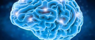
This is a hereditary disease that manifests itself as osteopenia and increased bone fragility. Osteogenesis imperfecta often runs in families and is associated with blue sclera, abnormal tooth development (dentinogenesis imperfecta), and progressive hearing loss.
Severe forms of the disease lead to the death of the fetus or newborn. Mild and moderate forms can occur in different ways. At birth there may be no deviations from the norm; the disease manifests itself later and progresses with age.
Many patients experience multiple fractures in infancy and childhood; during puberty, fractures occur less frequently, and then become more frequent again. In women, the risk of fractures is especially high during pregnancy and postmenopause.
Some women with mild disease do not develop fractures until postmenopause, in which case osteogenesis imperfecta is difficult to distinguish from postmenopausal osteoporosis. Classification. The classification developed by Sillens is most often used.
What is osteogenesis imperfecta?
Osteogenesis imperfecta (abbreviated OI or also called osteogenesis imperfecta , crystal man disease, Lobstein-Vrolik disease ) is a group of rare diseases that affect connective tissue and are characterized by extremely fragile bones that break easily, often for no apparent reason.
The specific symptoms and physical signs associated with the disease vary greatly from case to case. The severity of osteogenesis imperfecta also varies greatly, even among members of the same family. OI can be a mild disease or lead to severe complications. Four main types of OI have been identified. OI type I is the most common and mildest form of the disease. OI type II is the most severe disease.
In most cases, various forms of osteogenesis imperfecta are inherited in an autosomal dominant manner.
Definition of the concept
Osteogenesis imperfecta (from the Latin osteogenesis imperfecta ) is a genetic pathology of the human musculoskeletal system.
It is characterized by the fact that the baby’s bone structures become extremely fragile and susceptible to fractures at the slightest impact on them. This genetic disease is quite rare. Its main feature is that it is incurable.
“Crystal disease” occurs in both males and females. It occurs in one newborn baby out of 11,000 - 20,000 children born.
The first information about this pathology came in the seventeenth century.
A baby with this diagnosis may also have impaired growth, skin lesions, and sometimes muscle tissue, blood vessels, teeth, tendons and hearing organs are affected.
Although this genetic disease is incurable, there are methods that make life easier and normalize for children with Vrolik syndrome.
Signs and symptoms
With all types of osteogenesis imperfecta, associated symptoms vary greatly from case to case, even within families. Some affected people may experience no or only a few bone fractures; other victims may have multiple fractures. OI can range from a mild illness with few symptoms to a severe, debilitating disorder. The age at which fractures occur also varies from case to case.
— Osteogenesis type I.
Type I osteogenesis is the most common and usually mildest form of OI. In most cases, the disease is characterized by multiple bone fractures, which usually occur during childhood and puberty. Fractures usually begin when the affected child begins to walk; Fractures in the newborn (neonatal) period are rare. After puberty, the incidence of fractures usually decreases. Repeated fractures can cause minor deformities in the bones of the arms and legs (for example, bowed tibia and femur).
A distinctive feature associated with OI type I is a change in the color of the whites of the eyes to a bluish tint (blue sclera). In some cases, people with OI type I may develop pathologies of the middle and/or inner ear that contribute to or lead to hearing loss (for example, conductive and/or sensorineural hearing loss). Hearing loss most often occurs in the third decade of life; however, this can happen as early as the second decade or as early as the seventh decade.
People with OI type I may have a triangular face shape and an abnormally large head (macrocephaly). In about 50% of cases, people with osteogenesis imperfecta type I will experience a growth deficiency after birth (puerperium), resulting in little height (for example, affected individuals will be shorter than unaffected family members). About 20 percent of adults with OI type I develop an abnormal curvature of the spine from side to side or from front to back (scoliosis or kyphosis).
Additional symptoms associated with OI type I include loose (hyperextensible) joints, low muscle tone (hypotonia), and thin skin that bruises easily.
Some researchers believe that there is a subgroup of type I OI in which patients have dental pathologies in addition to the above-mentioned features.
— Osteogenesis type II.
OI type II is the most severe type of osteogenesis imperfecta. Affected infants often experience life-threatening complications at birth or shortly after birth. Infants with OI type II have low birth weight, abnormally short arms and legs (limbs), and bluish discoloration of the whites of the eyes (blue sclera). In addition, affected infants may have extremely fragile bones and numerous fractures at birth. The ribs and long bones in the legs of affected infants are often deformed.
Infants with OI type II often have underdeveloped lungs and an abnormally small upper chest, which can lead to life-threatening respiratory failure. In some cases, affected infants may experience congestive heart failure.
Infants with OI type II may have a small, narrow nose; small jaw (micrognathia); and an abnormally soft upper part of the skull (calvarium) with abnormally large soft spots. Affected infants may also have abnormally thin, fragile skin and low muscle tone (hypotonia).
Rehabilitation
Often, after the first few fractures, the parents of a child with osteogenesis imperfecta abandon an active lifestyle in favor of what they consider to be the safest, sedentary way of existence.
This reduces the effectiveness of drug treatment to zero and entails muscle atrophy and hypokinetic osteoporosis, which is much more prone to fractures.
Therapeutic exercise and massages are aimed at restoring joint mobility and the child’s ability to move independently.
A major role in rehabilitation is played by the psychological climate in the family, the motivation and social adaptation of the child.
Causes
In most cases, osteogenesis imperfecta types I, II and IV are inherited as autosomal dominant traits. Most cases of OI type II occur without a family history of the disease, but are the result of a spontaneous genetic change (ie, a new mutation).
Human traits, including classical genetic diseases, are the product of the interaction of two genes, one from the father and one from the mother. In dominant disorders, a single copy of the disease gene (received from the mother or father) will express itself as “dominant” over the other normal gene and result in the disease. The risk of transmitting the disease from an affected parent to offspring is 50 percent in each pregnancy, regardless of the sex of the child born. The risk is the same for every pregnancy.
Rare subgroups of OI types II and III can be inherited as autosomal recessive genetic traits. In recessive disorders, the condition does not occur unless the individual inherits the same defective gene for the same trait from each parent. If a person receives one normal gene and one disease gene, they will be a carrier of the disease, but usually not asymptomatic. The risk of transmitting the disease to a couple's children, both of whom are carriers of the recessive disease, is 25% in each pregnancy. The risk of having a carrier child, like the parents, is 50% with each pregnancy. The chance that a child will receive a normal copy of the gene from both parents and be genetically normal (for that particular trait) is 25%. The risk is the same for men and women.
Description of the pathology
Lobstein-Frolik disease is a genetically determined disease that occurs as a result of impaired bone formation. It leads to a decrease in bone mass and increased fragility. The pathology develops as a result of a defect in type 1 collagen, which is an important protein in the bone structure. Then it is produced in insufficient quantities or the structure of the substance is disturbed. For this reason, bones become weak and brittle. Because of this, the pathology is called “crystal disease.”
Reference. According to statistics, in approximately 50% of cases, imperfect bone formation is provoked by spontaneous mutations. The disease is diagnosed in 1 child out of 10–20 thousand newborns.
Crystal disease is incurable, but with the right approach it can make a child’s life much easier.
Disorders with similar symptoms
Symptoms of the following disorders may be similar to those of osteogenesis imperfecta. Comparisons can be useful for differential diagnosis:
- Achondroplasia is an inherited disorder characterized by abnormally short arms and legs and short stature (short limb dwarfism), abnormal facial features, and/or skeletal malformations. Characteristic facial features may include an abnormally large head (macrocephaly), an unusually prominent forehead, a low bridge of the nose, and/or underdevelopment of the midface (midfacial hypoplasia). Skeletal malformations may include unusually short fingers and toes (brachydactyly), an abnormally increased backward curvature of the spine (lordosis), outward bending of the legs, and/or narrowing (stenosis) of the spine. Additional pathologies may include limited elbow and hip extension, decreased muscle tone (hypotonia), and/or frequent middle ear infections (otitis media). In most cases, achondroplasia occurs accidentally, for no apparent reason (sporadic).
- Hypophosphatasia is a rare disease characterized by defective bone strengthening (mineralization), resulting in weakened bones. The long bones of the arms and legs may be too thick, short, and crooked. Affected infants may also have an abnormally small head (microcephaly). Many adults with hypophosphatasia are short in stature. Hypophosphatasia is inherited as an autosomal recessive trait.
- Pycnodysostosis is a rare disease characterized by increased bone density (osteosclerosis), short stature, an underdeveloped lower jaw, and dental disease. Patients often have fragile bones and may be susceptible to stress fractures. Pycnodysostosis is inherited as an autosomal recessive trait.
- Osteopetrosis is a rare disease characterized by increased bone density, brittle bones, and, in some cases, skeletal disorders. Although symptoms may not initially appear in people with mild forms of this disorder, trivial injuries can cause bone fractures due to bone abnormalities. Adult type osteopetrosis is milder than childhood malignant and intermediate osteopetrosis and can only be diagnosed in adolescence or adulthood when symptoms first appear. More serious complications occur with infantile malignant and intermediate osteopetrosis. Osteopetrosis can be inherited as an autosomal recessive or dominant trait.
- Osteogenesis imperfecta syndrome is a term used to describe a group of diseases characterized by bone pathologies (eg, brittle bones and multiple fractures) similar to those found in the main four types of OI. However, these disorders have associated features that distinguish them from the main four types of osteogenesis imperfecta. These so-called osteogenesis imperfecta syndromes are extremely rare, and only a few cases of each syndrome have been described in the medical literature. The Cole-Carpenter syndrome described below is osteogenesis imperfecta syndrome.
- Cole-Carpenter syndrome is an extremely rare disorder characterized by extremely fragile bones and multiple fractures similar to those seen in OI. In addition, affected infants may have blue sclera, premature closure of fibrous joints (cranial sutures) between some bones of the skull (craniosynostosis), an abnormally prominent forehead, an abnormally small jaw, and a protruding eye (proptosis). Affected infants may also experience growth retardation. There are about 5 cases of this disorder described in the medical literature. Cole-Carpenter syndrome is inherited in an autosomal dominant manner.
Etiology and pathogenesis
He. associated with disorders of protein or mineral metabolism, decreased osteoblast function or increased osteoclast activity. With O. n. note a normal or even increased number of osteoblasts and osteocytes with a normal number of osteoclasts, sufficient mineralization and pronounced basophilia) of the main substance of the bone, which indicates the normal development of the processes of resorption modeling of bone (see). A.V. Rusakov drew attention to the fact that the speech at O.N. This is not about changes in the quantitative composition of cellular elements, but about qualitative changes in their functions and activity. A large number of osteoblasts, which have high proliferative activity, produce little bone substance, quickly turning into osteocytes. With this O. n. differs from rickets, in which, on the contrary, a small number of osteoblasts produce significant masses of osteoid substance that do not undergo sufficient mineralization. The high proliferative activity of osteoblasts determines good healing of bone fractures in O. n. Thus, Ramser (LR Ramser, 1966), when examining a piece of tissue from the rib of a patient O. n. using a tetracycline label, he established that the rate of bone formation in this disease is three times higher than usual, but the transverse size of the rib remains half the normal size. This fact indicates that the fundamental possibility of growth of the bone skeleton during O. n. not broken. As a result of excessive compliance of the skull bones with O. n. they do not affect the growth of the organs contained in them, so the eyeball enlarges and the retina shines through the stretched and thinned sclera, giving it a blue tint; Due to hydrocele of the ventricles, the mass of the brain increases, which gives the head a characteristic spherical shape. The development of progressive deafness in O. was also explained by the disharmony of growth of the bone frame of the inner ear and the remaining bones of the skull. This explanation is based on the fact that with O. n. we are talking about local failure of the mesenchymal system of bone organs only. However, some researchers have shown that the inferiority of the mesenchyme is of a more generalized nature and is manifested by weak production of collagen by fibroblasts of the skin, ligaments, membranes of the eyeball, etc. This position makes it possible to combine a large group of syndromes and diseases, which are based on a common genetic defect of the mesenchyme with a predominant lesion in each syndrome of one or another of its segments - osteogenesis imperfecta, desmogenesis imperfecta (see), chondrogenesis imperfecta (see), Marfan syndrome (see). Weak formation of dentin in the tooth buds during O. n. explain, for example, the imperfect development of teeth, which are very quickly susceptible to caries.
Modern studies have shown that with O. n. there is insufficient production of collagen (see). With O. n. Predominantly precollagen fibers are produced, which do not undergo maturation, or collagen of a special qualitative composition. Histochemical studies indicate that collagen fibers in O. n. have an unusually high proline content and that such collagen inhibits calcification processes in vitro, although the light-optically determined processes of bone mineralization during O. n. remain unchanged.
Diagnostics
The diagnosis of osteogenesis imperfecta is made based on a thorough clinical assessment, a detailed patient history, identification of characteristic symptoms and various specialized tests. Surgical removal and microscopic examination (biopsy) of the skin may be performed to determine the presence of collagen abnormalities. A blood sample may be taken and tested to identify the genetic mutation that causes OI.
In some cases, the diagnosis of OI can be made before birth (prenatally) based on specialized tests such as ultrasound, amniocentesis, and/or chorionic villus sampling. Ultrasound examinations may reveal telltale signs such as fractures or bowed long bones. During amniocentesis, a sample of the fluid surrounding the developing fetus is removed and examined. During chorionic villus sampling, a tissue sample is taken from part of the placenta. Chromosomal studies performed on this fluid or tissue sample may reveal the genetic mutation causing osteogenesis.
Standard Treatments
Treatment for osteogenesis imperfecta is aimed at eliminating the specific symptoms that occur in each person. Treatment is aimed at preventing symptoms, maintaining individual mobility, and strengthening bones and muscles.
Exercise and physical therapy programs have proven effective in strengthening muscles, increasing stress-bearing capacity, and reducing susceptibility to fractures. Physiotherapy in water (hydrotherapy) has also been found to be helpful, as movement in the water reduces the likelihood of fracture. Individuals with OI should consult their doctors and physical therapists to determine a safe and appropriate exercise program.
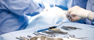
People with OI often use a procedure in which metal rods are surgically placed in long bones to prevent fractures. Plastic braces are replacing plaster casts as protective devices because they allow greater freedom of movement and can be used in water. Inflatable suits can provide additional protection, especially for very young children.
Surgery may be necessary for people with severe spinal bone deformities or basilar intussusception. Dental procedures may be required to correct various dental abnormalities. Patients, especially adults, should be evaluated for hearing impairment, often associated with OI. Other treatments are symptomatic and supportive. Genetic counseling is recommended for families affected by this disorder.
Treatment of “crystal children”
Due to the fact that Vrolik syndrome is a genetic pathology, therapy will be only symptomatic. That is, it will be aimed at improving bone mineralization, preventing fractures and rehabilitation measures.
A baby with “crystal disease” needs a gentle regimen, and also requires therapeutic and gymnastic exercises, massages, hydrotherapy, and physiotherapeutic procedures. Among the medications, a child with this pathology is prescribed multivitamins, vitamin D, and hormonal medications (containing growth hormone).
To increase collagen synthesis, somatotropin is prescribed.
An improvement in the clinical picture can be achieved by administering biophosphates, as they inhibit the destruction of bone structures.
Fractures require careful repositioning of the bones and plaster immobilization. If a child has severe deformation of bone structures, then he needs surgical treatment. Such children undergo corrective osteotomy and intramedullary or periosteal osteosynthesis.