Glaucoma occurs against a background of persistently elevated intraocular pressure. This is a chronic ophthalmological disease with serious complications - narrowing of the visual field and damage to the optic nerve. Glaucoma cannot be completely cured; at best, it is only possible to maintain visual functions at a relatively acceptable level. Let's look at the causes of glaucoma, methods of prevention and reduction of intraocular pressure.
Signs of increased intraocular pressure
The visual organs are a hydrodynamic system. Inside the eyeball there is fluid under pressure, which is regulated by the inflow and outflow system. If the outflow of intraocular fluid does not occur on time, increased pressure is created. Also, increased ophthalmotonus is created by excessive production of intraocular fluid.
The intraocular moisture is saturated with beneficial substances that nourish the inner corneal layer and lens. The outer side of the cornea receives nutrients from the tear fluid. Also, the intraocular fluid takes part in the metabolic process inside the visual structures: it eliminates protein breakdown products and carbon dioxide.
People at risk for increased intraocular pressure include those suffering from:
- high blood pressure;
- severe myopia;
- renal failure;
- diabetes mellitus;
- pathologies of the endocrine system.
Also at risk are people exposed to constant stress and women during menopause. Intoxication with chemicals also has a detrimental effect on the circulation of fluid inside the eye chambers.
Increased ophthalmotonus
Signs of increased ophthalmotonus:
- constant headaches in the temporal and suprafrontal region;
- discomfort and heaviness in the eyes;
- rapid fatigue with light visual loads;
- the appearance of spots before the eyes;
- redness of the sclera (but not always).
A characteristic sensation for increased ophthalmotonus is a feeling of fullness inside the eyes. This is especially noticeable when you lightly press your fingers on closed eyelids. Patients also note a narrowing of the field of vision, blurred visualization in the twilight, blurred objects, and blurred vision. With increased ophthalmotonus, lateral vision disappears, and sometimes attacks of glaucoma are accompanied by nausea.
Patients often attribute visual fatigue to fatigue and try to restore visual function with good rest. But that doesn't help. If discomfort in the eyes occurs, it is necessary to undergo an examination by an ophthalmologist, especially for patients over forty years of age.
The insidiousness of glaucoma is the long-term absence of any symptoms of increased pressure. Pathology can be detected at an early stage only using hardware examination methods. Therefore, do not delay your visit to the ophthalmologist if you feel even slight discomfort in the visual organs or a decrease in the quality of vision.
How to prevent vision loss from glaucoma
If there is a family history of glaucoma, it should be checked systematically, especially after 35 years. If there are risk factors, intraocular pressure should be measured annually or even more often. People whose profession or lifestyle involves visual stress should visit a doctor at least 2 times a year.
At an early stage of the development of glaucoma, the patient is prescribed drip administration of antihypertensive antiglaucoma drugs. If drop therapy is sufficient to stabilize the pressure, the patient is sent to be observed by an ophthalmologist. It is prohibited to cancel or replace medications prescribed by a doctor, as well as change the dosage, time and frequency of instillation. Many patients with glaucoma use antihypertensive drops for life.
If conservative therapy does not help normalize pressure at the initial stage of glaucoma, the patient is recommended laser treatment. A simple operation using a laser helps improve the functionality of the drainage system and improve the outflow of intraocular fluid. Laser treatment of glaucoma is painless, the operation is performed quickly and on an outpatient basis. This method has its contraindications.
Indications for microsurgery for glaucoma:
- conservative treatment is ineffective;
- laser surgery did not help normalize blood pressure;
- the disease was diagnosed at a late stage of development;
- there is severe damage to the structures of the eye;
- severe pain syndrome.
Microsurgery is the most effective method of treating glaucoma.
Recommendations for patients with glaucoma
- Avoid taking atropine, belladonna, strychnine, caffeine;
- Glaucoma should be reported to every doctor who prescribes treatment for concomitant diseases in any area;
- treatment of concomitant disorders is mandatory;
- it is necessary to consult an ophthalmologist regarding the use of any medications;
- it is important to maintain normal functioning of the gastrointestinal tract (timely treatment of constipation);
- with angle-closure glaucoma, sudden changes in lighting should not be allowed;
- before visiting a cinema or other dark room, it is advisable to instill pilocarpine;
- Don’t overwork yourself;
- avoid any physical and nervous strain;
- with glaucoma, you cannot work in an inclined position;
- You can read and watch TV only in good lighting;
- Every hour of concentrated visual work you need to take a break for 10 minutes;
- It is necessary to choose a diet according to age;
- It is useful to limit the amount of animal fats and sugar in the diet;
- It is healthy to eat raw vegetables and fruits, lean meat and boiled fish (no smoked or salted meat);
- if there are no indications, the amount of liquid is not limited, but it is not recommended to drink more than a glass at a time;
- with glaucoma, it is dangerous to smoke, abuse alcohol, natural coffee and strong tea;
- It is undesirable to wear tight ties and collars, they interfere with blood circulation in the neck and head;
- you need to sleep on a high pillow for 8 hours;
- A moderately active lifestyle is important (gymnastics, walks in the fresh air);
- hypothermia must be avoided; you should not swim in cold waters even in warm weather;
- protect your head from the sun;
- refuse to visit the sauna, bathhouse and solarium.
The disease develops without pronounced symptoms, so people often seek help when glaucoma has already affected the retina and nerves. If you can lower the pressure, then you can no longer reverse the damage.
Sources used:
- Textbook of eye diseases / V.N. Arkhangelsky, M.K. Bryantseva, K.V. Dormidontova. - M.: State Publishing House of Medical Literature, 1983.
- Ultrasound in ophthalmology / Diana Angelova. - M.: Palmarium Academic Publishing, 2012.
- Age-related macular degeneration / A.S. Alpatov. - M.: GEOTAR-Media, 2015.
- Department of Ophthalmology and Visual Sciences University of Wisconsin System
Types of glaucoma
Pathology of intraocular pressure is observed at any age. In this regard, there are several types of glaucoma:
- congenital;
- infantile;
- juvenile;
- an adult.
Congenital pathology differs from genetic pathology in that it was formed during the intrauterine development of the fetus. Also considered congenital are pathologies that appear during the passage of the fetus through the mother’s birth canal. During the period of intrauterine formation of the fetus, various pathologies may appear caused by abnormal development. If by the time of birth the embryonic tissues of the fetus do not dissolve, the child is born with a pathology of the circulation of intraocular fluid. During the birth process, the fetus also faces many dangers: dislocation of the lens, infection of the choroid with further inflammation, etc.
Congenital glaucoma can manifest itself either immediately after childbirth or at any other time. Congenital glaucoma itself manifests itself during the first three years of a child’s life, infantile glaucoma manifests itself during the first ten years, and juvenile glaucoma after 10 years.
In addition, glaucoma is distinguished:
- primary;
- and secondary.
Primary
Primary is formed due to impaired circulation of intraocular fluid. This pathology develops independently and is not caused by other ophthalmological diseases. Primary glaucoma can be congenital (genetically predisposed), which appears due to pathology of the nervous regulation of the functions of the optical apparatus, which appears due to age-related changes in tissues or disorders of blood circulation and metabolic processes.
Blockage of the excretory passages may also be due to excessive production of pigment (coloring substance) of the iris. In this case, pigment cells accumulate in the iris, preventing moisture circulation.
Secondary
Secondary glaucoma is formed due to external causes:
- as a consequence of ophthalmological operations;
- as a consequence of injury to the visual organs;
- as a consequence of infection of the visual organs.
Secondary glaucoma always develops against the background of existing pathologies of the visual organs. When the causes that caused it are eliminated, the pathology completely disappears. However, the causes of increased ophthalmotonus must be eliminated before irreversible changes develop within the ocular structures.
In infectious diseases of the eyes that affect the choroid, adhesions are formed. They create obstacles to the correct circulation of intraocular fluid. Sometimes the result of infectious processes is a congestion of the pupil, which also interferes with the inflow/outflow of moisture.
When the lens is deformed (trauma, dislocation, cataract), the circulation of fluid inside the eye chambers is also disrupted, which is accompanied by an increase in pressure. Damage to the network of blood vessels limits the flow of nutrients into the retina, which provokes the formation of new vessels to compensate for nutritional deficiencies. The network of new vessels clogs the ducts, and fluid circulation becomes impossible or malfunctions.
Burns of the eye mucosa often provoke an increase in ophthalmotonus due to the formation of adhesions and connective scars that remain after the integrity of the eyeball mucosa is restored. Scars and adhesions become a natural obstacle to the complete outflow of fluid. For the same reason, secondary glaucoma may appear after ophthalmic surgery on the eyeball.
Another cause of glaucoma is the absence of a lens. This can be either a congenital pathology or acquired - after surgery to remove the lens body. In this case, the vitreous body shifts, which causes a disruption in the circulation of intraocular fluid.
Neoplasms in the orbit or posterior chamber of the eyeball also cause obstacles to hydrocirculation, blocking the outflow angle.
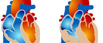
Closed and open angle
If the disease is caused by incorrect outflow of intraocular fluid, we speak of glaucoma:
- closed angle;
- open angle.
The word “angle” in the diagnosis refers to the plane between the iris and the inner surface of the corneal layer. Through this angle, intraocular moisture circulates. Angle-closure glaucoma is the most dangerous form, as it forms suddenly and causes severe pain. It is the closed-angle form that in most cases ends in loss of visual functions.
The open-angle form of glaucoma develops gradually, without causing pronounced symptoms. Over a period of time, the angle of the anterior chamber of the eye undergoes pathological changes, as a result of which correct fluid circulation becomes impossible.
What is mixed glaucoma? This disease is characterized by open-angle symptoms and closed-angle attacks. This is a very difficult type of disease to treat, requiring an integrated approach.
However, increased intraocular pressure is not unique to glaucoma. It is often a sign of intraocular hypertension, in which the quality of vision does not suffer. In order not to worry about your eye health, you need to consult an ophthalmologist. Only a specialist has the right to make a true diagnosis.
Drugs used in therapy
Drops used to enhance the outflow of fluid inside the eye, narrowing the pupil and retracting the iris, which allows excess fluid to drain and thereby reduce pressure:
- Xalatan,
- Oftan
- Polycarpine,
- Travatan,
- Polycarpine hydrochloride.
A medicine of the same effect, which, among other things, is prescribed for corneal dystrophy, as well as for bronchial asthma, bradyarrhythmia, dry keratitis:
- Arutimol,
- Azopt,
- Niolol,
- Betoptik,
- Cusimolol,
- Timolol.
Combined drugs intended for the treatment of glaucoma, the purpose of which is to reduce the aqueous fraction of intraocular contents and improve their outflow:
- Fotil-forte,
- Fotil,
- Timpilo.
And the use of a drug such as Simax (methionyl-glutamyl-histidyl-phenylalanyl-prolyl-glycyl-proline), which is intranasal drops that quickly reach the brain tissue, which is very important for pathological changes inside the eye.
There may be metabolic failures in the tissues of the eye, which are especially typical for elderly patients, therefore, in addition to direct-acting medications, drugs are also prescribed that increase overall tone and strengthen the immune system. Such as ATP (adenosine triphosphoric acid), cocarboxylase, B vitamins and vitamin E.
Treatment of glaucoma without surgery requires compliance with certain conditions. This is a mandatory agreement with the attending physician on the list of absolutely all medications used.
If a patient has a disease of the musculoskeletal system and is prescribed chondroprotectors, then some drugs on the ophthalmologist’s list will either be contraindicated or cause side effects. The same applies to medications that help with diseases of the gastrointestinal tract.
Causes of glaucoma development
Most often, the disease occurs against the background of a genetic predisposition, with the exception of traumatic factors (surgeries, mechanical damage, infections). However, the following reasons contribute to the development of the disease:
- age-related changes in the tissues of the visual organs;
- severe ophthalmological diseases;
- diseases of the endocrine system (including obesity);
- cervical osteochondrosis.
Open-angle glaucoma is formed due to degenerative age-related changes in the tissues of the optical apparatus (sclerotic plaques on blood vessels, cataracts, high blood pressure). However, age-related changes do not always become the cause of this pathology; the danger of increased ophthalmotonus also appears with severe myopia. High myopia is characterized by excessive production of intraocular moisture, which complicates its outflow.
The closed-angle shape is characteristic of farsightedness, in which the shape of the lens is constantly enlarged. In this case, there is a sharp closure of the angle of the anterior chamber of the eye, which is responsible for the circulation of moisture. If myopia is characterized by an enlarged shape of the eyeball, then with farsightedness its diameter decreases (and the size of the lens increases). Pathological changes in the shape of the eyeball and lens with the cornea play a leading role in the formation of increased ophthalmotonus.
What factors can trigger a sudden attack of angle-closure glaucoma? Pain may occur with:
- long stay in a dimly lit room;
- excessive fluid intake;
- using medications to dilate the pupil;
- when tilting the head down for a long time.
Angle-closure glaucoma also occurs due to chronic stress, which causes the iris to shift toward the lens. An attack can also be triggered by displacement of the vitreous body due to the appearance of a tumor in the optical apparatus. The formation of a tumor pushes the lens forward, thereby closing the anterior chamber angle.
Glaucoma without pressure
Glaucoma can also develop against the background of normal intraocular pressure. In this case, there is a gradual narrowing of the field of vision, pathological changes in the optic nerve and loss of visual functions. This is an extremely rare type of pathology, the causes of which are unknown to medicine. Disruption of the blood supply to the retina, the development of atherosclerosis and too high intracranial pressure can lead to the development of the disease.
Acute closed-angle shape
In acute angle-closure form, the patient has:
- Severe headache (usually a pressing pain in the parietal and temporal regions)
- Pain in the eyes
- Nausea and vomiting (occur as a result of nervous tension from long-lasting headaches)
- Blurred vision
- Appearance of a halo around light sources
- Redness of the eyes
These symptoms are not present all the time; they may occur for one to two hours, then subside and subsequently reappear. But with each new occurrence, visual function is damaged. This damage does not recover, so it is necessary to begin treatment as soon as possible.
Symptoms of glaucoma
It was already noted above that glaucoma is not characterized by pronounced symptoms. The closed-angle form manifests itself during an attack - suddenly, with severe pain. At first, the pain affects only the eye, then spreads to the entire half of the head. When palpated, the eye feels hard like a stone. During an attack, the affected eye becomes bloodshot, and the patient begins to feel very sick, even to the point of vomiting. Often there is a slowdown in heart rate, accompanied by pain in the heart area.
It is necessary to urgently call an ambulance, otherwise you may completely lose your vision. Painkillers will not help preserve the optic nerve, which suffers during a severe attack. An attack of angle-closure glaucoma is often confused with a hypertensive crisis, which has similar symptoms. It is necessary to check the condition of the eyeball: with glaucoma, it becomes stony to the touch and very painful when touched.
The open-angle form does not manifest itself for a long time; glaucoma is most often discovered by chance during an examination by an ophthalmologist. Some signs that indirectly indicate a problem with the visual apparatus:
- excessive moisture in the eye mucosa;
- rapid fatigue during visual stress;
- frequent pain in the temporal/frontal region not associated with a cold;
- double vision of objects;
- blurred vision;
- blurred visualization at dusk or in poor lighting;
- the appearance of multi-colored circles when observing a light source;
- narrowing the field of view.
The gradual deterioration of vision, accompanied by the listed symptoms, should alert everyone. If measures are not taken to preserve visual functions, the narrowing of the field of vision will become catastrophic: a person sees the world as if through a spyglass.
Note! A distinctive feature of glaucoma is precisely the narrowing of the visual field. The clarity of visualization is not impaired if there is no myopia.
The patient is surprised to notice that he no longer sees the tip of his own nose. This indicates a lack of peripheral vision, for which the optic nerve is responsible. There may be other variations in the narrowing of the field of visibility: the patient sees the tip of the nose, but does not notice its profile (back). As the pathology develops, the patient stops seeing objects on the sides, as if they do not exist. This gradually narrows the field of vision, leading to a complete loss of visual functions.
People under forty years of age are not at risk of developing glaucoma; the chance of getting the disease increases sharply after 50 years of age.
Prevention
Source: moiglaza.com
In principle, anyone can get glaucoma without noticing it in time. Therefore, only preventive examinations by an ophthalmologist can protect against the development of this disease.
Unfortunately, this is still done too rarely because the public is not sufficiently informed about the dangers of developing glaucoma.
Preventive examinations become even more important if you remember that the damage caused by glaucoma, even after surgery, will remain irreversible.
The purpose of surgery for glaucoma can only be to preserve the existing visual ability and prevent the onset of blindness. Preventive examinations include all examinations that are carried out to diagnose the disease, and are described below in the corresponding passage.
In cases where surgery is performed by opening the eyeball, it is almost impossible to avoid complications, but it is still necessary to prevent their occurrence.
In most cases, the preventive component of treatment depends on the professionalism of the doctors under whose supervision the patient is.
Also, a lot depends on the equipment with which the surgical intervention will be carried out, as well as on the quality of the consumables involved in the operation.
From this we can conclude that the prevention of complications depends on the following factors:
- professionalism of an ophthalmologist;
- equipment on which the operation is performed;
- quality of consumables.
Of course, a lot depends on how advanced the disease was, because the more complicated the operation, the more likely it is that complications will arise after it. Prevention of complications is a very important factor in minimizing their manifestations
For whom is it especially important to undergo preventive examinations?
Preventative screenings are especially important for people with risk factors for glaucoma.
These include people over 40 years of age who have a history of this disease in their family, people with myopia (with diopters higher than -5), people with diabetes with significant changes in the fundus of the eye, and people with circulatory disorders.
If the first examination does not reveal any symptoms of glaucoma, the next examination can be carried out after 3 years, if your age does not exceed 65 years. Older people over this age should be checked every 1-2 years, even if the first examination did not reveal any abnormalities.
Diagnostic methods
How is an eye examination for glaucoma performed? Experienced ophthalmologists determine ophthalmotonus by palpation, but few doctors are capable of this. Most often, the disease is diagnosed using hardware methods - old and new. Modern clinics are equipped with the latest equipment for fundus examination.
Examination of ophthalmotonus is carried out using:
- Maklakov tonometer;
- electrotonography;
- pneumotonometry;
- pachymetry.
Maklakov tonometer is an old method for determining ophthalmotonus. First, the patient is instilled with a medicine that causes a temporary spasm of accommodation. This is necessary to stop the reaction of the lens to near/far objects. Then colored weights are placed on the cornea. Based on the condition of the weights, the ophthalmologist determines ophthalmotonus.
Electrotonography is a modern non-contact method for studying intraocular pressure and the quality of the hydrodynamics of the optical apparatus. Examination of the state of the visual organs with a pneumotonometer does not give a convincing result, so additional devices are used to clarify the diagnosis.
Pachymetry allows you to examine the thickness of the corneal layer. With thickening of the cornea, ophialmotonus is actually lower than the indicators of the devices, and with a thin cornea, it is higher than the indicators. Diagnostics is used to clarify hardware data. Patients are also examined using perimetry - identifying dark spots in the field of view. Many patients do not even know about them.
In what case are we talking about pathology of intraocular pressure? The indicators depend on the type of device used. On average, pressure up to 22 mmHg is considered normal, but there may be deviations.
Examination of the angle of the anterior chamber of the eye:
- gonioscopy;
- biometroscopy;
- tonography;
- ophthalmoscopy;
- other methods.
The functionality of the anterior chamber angle is determined by gonioscopy and biomicroscopy. The depth of the anterior chamber is examined, which allows you to determine the likelihood of developing glaucoma. Tonography examines the production and quality of circulation of intraocular fluid. Ophthalmoscopy examines the condition of the optic nerve in detail by dilating the pupil. For clarifying diagnostics, other hardware methods for examining the optical apparatus may be prescribed.
A single diagnosis of the fundus and the level of ophthalmotonus does not provide a comprehensive answer, so the diagnosis is carried out several times in a row.
Making a diagnosis is complicated by the structural features of the patient’s body. Each person has his own threshold for exceeding normal intraocular pressure, as well as the sensitivity of the optic nerve to pressure.
Folk remedies
Is glaucoma treated at an early stage using folk remedies? These prescriptions are not as effective as medications, but can slow down the progression of the disease if diagnosed at an early stage.
Here are some recipes:
- Dry calendula flowers pour 300 ml of boiling water. Leave for 5-6 hours in a dark place, strain through cheesecloth. Wipe your eyes with the decoction in the morning and evening.
- Dilute liquid honey with boiled water in a ratio of 1:3. Place 1-2 drops into eyes twice a day.
- Dilute freshly squeezed aloe leaf juice with boiled cool water in equal parts. Place 2-3 drops into the eyes once a day.
It is also recommended to make compresses from raw grated potatoes or tea leaves.
These recipes are a kind of prevention of eye pathologies. If such treatment is not effective, you should contact a qualified specialist.
Methods of drug therapy
Medicines are prescribed according to the reasons that caused the increase in intraocular pressure. This is either uncontrolled production of intraocular fluid or a violation of its outflow. Along with targeted drugs, medications that improve metabolic processes in tissues and vitamins for the eyes are prescribed.
The choice of medications is the responsibility of the ophthalmologist. It is strictly forbidden to prescribe treatment on your own, since unproductive therapy will lead to partial or complete loss of visual functions. The patient must undergo constant examination, scrupulously follow the ophthalmologist’s recommendations and not change the frequency of medication intake according to his own judgment. It is very dangerous.
Drugs to reduce ophthalmotonus have many side effects. It is necessary to discuss in advance with an ophthalmologist the use of analogues or generics in case of intolerance to the prescribed medication. Any discomfort when using eye drops should be reported to your doctor.
You should not neglect taking vitamin complexes to improve visual functions. These drugs help restore metabolic processes, blood circulation, and nutrient deficiency in tissues. With vitamins, visual functions will be restored faster than without them. Vitamins stop the development of age-related degenerative processes in tissues, remove toxic substances and protect against the negative effects of environmental factors (UV rays, smoke, poor ecology, dry air, dust, etc.).
How to prevent negative consequences?
Early diagnosis of the disease ensures effective treatment.
To avoid serious consequences, it is important to notice the increase in intraocular pressure in time and conduct a comprehensive diagnosis of the disease. To do this, you need to regularly visit an ophthalmologist for preventive examinations.
It is enough for young people to do this once every few years, after 40 years - every year, after 60 years and people who are at risk, especially due to several factors simultaneously, should visit a doctor twice a year. The following methods are used to diagnose the disease:
- perimetry;
- biomicroscopy;
- tonometry;
- ophthalmoscopy;
- gonioscopy.
Does it still seem difficult to heal?
It has been established that 20-30% of the adult population suffer from the problem of pressure surges. With age, the prevalence of the disease increases and reaches 50-65%. The consequences of high blood pressure are known to everyone: these are irreversible damage to various organs (heart, brain, kidneys, blood vessels, fundus of the eye).
In later stages, coordination is impaired, weakness appears in the arms and legs, vision deteriorates, memory and intelligence are significantly reduced, and a stroke can be triggered.
It is impossible to completely get rid of the disease. Therapy is aimed at lowering intraocular pressure, preventing or stopping the process of destruction of the optic nerve, stopping the progression of the disease and its negative consequences. Various methods are used to treat glaucoma:
- medicinal;
- laser;
- surgical.
The therapeutic method, drugs and dosages are selected individually in a particular case. You cannot self-medicate, as it is ineffective, and the lack of qualified medical care can cause complications.
In many cases, medications can delay the progression of the disease. Taking them throughout life, a person manages to preserve his vision. Subject to the recommendations of the ophthalmologist and proper therapy, the patient can lead a normal lifestyle.
You can eat caffeine-containing products, exercise, visit the sauna and steam bath, and read in the right lighting. It is worth getting more rest and maintaining a sleep schedule, and you should avoid excessive drinking, smoking and wearing clothes that restrict blood circulation in the neck.
Laser surgery
Some forms of glaucoma can only be treated with surgery. Modern advanced surgery makes extensive use of laser treatment.
Laser trabeculoplasty is used to clear blocked open-angle ducts. The surgery is performed under local anesthesia (eye drops). An optical lens is placed in front of the patient's eye through which the laser beam is passed. Thanks to this correction, the outflow of intraocular moisture occurs unhindered.
To reduce moisture production, another correction method is used - cycodiodic. The laser beam precisely destroys the area where intraocular moisture is produced, thereby reducing its amount. Optimizing fluid production automatically reduces ophthalmotonus.
Iridectomy is another method of minimally invasive intervention in the structures of the visual organs. The operation is used to relieve an acute attack of angle-closure glaucoma. A laser beam excises a certain area of the iris, which opens access for the correct circulation of intraocular moisture.
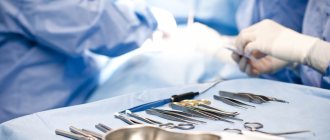
Traditional surgery
Sometimes drug therapy and laser do not cope with the task. In this case, traditional methods of invasive intervention - trabeculectomy - come to the rescue. The operation is indicated for any type of glaucoma. The method is based on making incisions on the sclera to drain excess moisture. There is no need to fear that all the intraocular fluid will leak out through the incisions: they are microscopic enough for this. The moisture will gradually drain into the conjunctival sac, relieving the eye chambers of pressure. If the drainage system becomes overgrown, notches will have to be applied again.
iStent
This innovative method has been used relatively recently, but has proven itself to be the best. The iStent device is a microchip implanted into the lining of the eye. It allows excess fluid to leave the eyeball as it fills, thereby stabilizing the internal pressure in the chambers. The microchip is absolutely not felt by patients and does not interfere with everyday life.
The advantage over traditional notches on the sclera is obvious: the tissue remains intact, without cuts. The disadvantage of this method is that it cannot be used for angle-closure glaucoma. A microchip can be used at any age and is safe for health. Mini iStent is the smallest medical device ever used in medical practice.
Postoperative treatment
An ointment bandage is applied to the operated eye. It is important not to rub the operated eye under any circumstances or apply mechanical pressure to it.
In addition, in the first days after surgery, you should not overstrain the operated eye by reading or watching TV for a long time. Also, for the first time after cataract surgery, you should avoid physical activity and not go to the sauna.
Complications
Although surgery for glaucoma is a safe treatment, complications cannot be completely ruled out. The repeated increase in intraocular pressure is usually only temporary.
Bleeding in the anterior eye chamber, although it looks dramatic, requires, however, only in special cases, as an exception, medical intervention. The complications listed below are so rare that they are not even included in the statistics.
However, they are listed here for the sake of completeness of information: when local anesthesia of the affected eye by injection, in rare cases, seizures, bleeding in the eyelids and eye orbit, as well as blindness may occur.
Consequences of the operation
Antiglaucoma surgery is considered a rather complex operation and sometimes leads to complications. Thus, having undergone surgical intervention, as a result it may simply not give positive dynamics and the course of the disease will only worsen.
Intraocular pressure will remain high, but the patient himself is unlikely to be able to measure it at home, so in this case, the complication is that the disease continues to progress, which can have a rather negative impact on eye health.
Also, a patient who has undergone surgery may experience a veil that appears and disappears, fuzzy halos appear around light sources, and the eyes and temples may hurt - this indicates an increase in intraocular pressure.
Bottom line
The death of the optic nerve cannot be cured; this process is irreversible. Therefore, to preserve visual functions, it is important to promptly detect the development of glaucoma. It is impossible to independently detect increased ophthalmotonus; it is determined only through hardware diagnostics. After forty years, it is very important to undergo a preventive examination by an ophthalmologist for increased intraocular pressure.
To prevent problems with circulation and the production of intraocular fluid, it is necessary to perform simple eye exercises that relieve tension and improve blood circulation. It is especially important to do eye exercises for people who work at computers. Constant overstrain of the visual apparatus leads to adverse consequences even at a young age.
Also, to prevent any ophthalmological diseases, it is useful to take vitamin complexes formulated specifically for eye health. They can be selected with the help of an ophthalmologist, in accordance with identified problems or to prevent them. We should not forget about foods that are healthy for the eyes, especially blueberries.
Sources used:
- Lesions of the nervous system and organ of vision / R.I. Korovenkov, L.M. Tibekina. - M.: ELBI-SPb, 2012.
- Diagnostic reference book for an ophthalmologist / P.A. Bezdetko, S.F. Zubarev, N.V. Panchenko. - M.: Phoenix, 2006.
- 100% vision / S.V. Dubrovskaya. - M.: Book on Demand, 2009.
- Wikipedia article
Adaptation to medications and the need to replace them
Before starting a course of medication, the doctor will definitely conduct diagnostic tests for the entire range of medications that he deems necessary to use. And after starting treatment, constant dynamic monitoring of the condition of the eyes is carried out. Monitoring is carried out daily or every other day or two during the first two weeks of using the prescribed drugs.
In the future, given the duration of treatment for glaucoma, it is necessary to consult and be examined by specialists at least two to three times a month. After possible detection of resistance to the selected drug and after dynamic tests, the drug is replaced.
Given the abundance of drugs on the ophthalmological market, there is practically no problem of addiction to one or another drug - their diversity allows you to vary their use.