General information
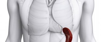
The spleen is an unpaired organ located in the abdominal cavity.
Its size increases with age. In adults, the weight of the spleen is 150-170 g. Normally, the spleen is hidden by the lower ribs and is not palpable. It can be detected by palpation with a magnification of 1.5-3 times. It is a well-supplied lymphoid organ that performs various functions. Functions of the spleen in the body:
- Participation in the formation of an immune response to foreign antigens. This organ is involved in the production of antibodies (synthesis of IgG , properdin and tuftsin ).
- Filtration of blood and elimination (removal) of microorganisms and blood elements loaded with antibodies. This organ is a large accumulation of reticuloendothelial tissue, the main function of which is phagocytosis performed by macrophage cells. Normal and abnormal blood cells are also eliminated.
- Participation in lymphopoiesis. The white pulp of the spleen, as a lymphatic organ, produces lymphocytes .
- Depositing blood. This function is performed by the red pulp of the spleen. Normally, one third of all platelets and a large number of neutrophils, which are produced during bleeding or infection, are deposited in it.
- Participation in hematopoiesis in certain diseases (in adults, the organ of hematopoiesis is the bone marrow).
- Regulation of portal blood flow due to the common vascular network with the liver.
- Participation in metabolic processes.
In many conditions and diseases, changes in the spleen in the form of splenomegaly are observed. Splenomegaly, what is it? This term is used to refer to the enlargement of a given organ to one degree or another. A synonym is the term megasplenia. Changes in size and structure occur in response to infectious, immune and hemodynamic factors. Changes in the spleen always indicate an acute or chronic process in the body - this is an indicator of pathological conditions.
Splenomegaly is an early and sometimes leading symptom of a number of diseases, without being an independent nosological entity. You need to know that an increase in this organ is always the body’s reaction to hematological, non-hematological diseases or a consequence of portal hemodynamic disorders. Therefore, the classification always takes into account the underlying disease against which this symptom developed. The splenomegaly code according to ICD-10 R16.1 is established if the enlargement of this organ is not classified in other categories.
Splenomegaly is often, but not always, accompanied by hypersplenism . Hypersplenism is a pathological condition that is characterized by a decrease in the number of formed elements (erythrocytes, leukocytes, platelets) in the circulating blood due to increased spleen function. pancytopenia develops , which is life-threatening.
Hypersplenism is associated with increased blood-destructive function of the spleen. This may be due to a number of reasons: difficulty in the outflow of blood from this organ, increased contact of formed elements with spleen macrophages, or increased activity of macrophages. Changes appear only in the peripheral blood, and the bone marrow remains normal. In many cases, the spleen is enlarged without symptoms of hypersplenism , and the latter can also occur with normal spleen sizes.
In most patients, hypersplenism requires removal of the spleen, after which the blood picture and the patient’s condition return to normal.
Prevalence
Normally, the spleen cannot be felt during palpation. Statistical studies on the topic of splenomegaly in the USA have shown that in practice, the spleen can be palpated, according to various sources, in 2–5% of the population.
It is believed that splenomegaly affects representatives of all races equally. However, in black residents of malaria-endemic countries, an enlarged spleen may also be caused by the presence of mutant hemoglobins S and C in the blood.
A separate issue is tropical splenomegaly - an enlargement of the spleen, which sometimes occurs in tourists who visit African countries, and women are susceptible to it twice as often as men.
Pathogenesis
With splenomegaly of various etiologies, different changes occur; therefore, several mechanisms of this symptom are considered:
- In response to antigenic stimulation during bacterial and viral infections, with a large number of abnormal cells during hemolysis hyperplasia of its reticuloendothelial tissue develops Hyperplasia of lymphoid tissue also occurs as antibody synthesis increases. All these mechanisms are associated with an increase in the protective function - “working splenomegaly”.
- An increase in size against the background of abnormal blood flow in the organ is observed with blood stagnation due to increased pressure in the portal vein or thrombosis of the splenic vein. In both cases, the outflow of venous blood from the spleen becomes difficult. Overflow of blood causes hyperplasia of the connective tissue, and it increases. The spleen compensates for the increased pressure in the portal vein by taking on the increased load. Splenomegaly during congestion occurs with hyperplasia of the reticular tissue and proliferation of connective tissue. The degree of organ enlargement depends on the level of blockage - extrahepatic (or subhepatic, which is associated with pathology of the portal vein vessels), intrahepatic (associated with cirrhosis ) or suprahepatic ( hepatic vein abnormalities ). subhepatic portal hypertension occurs , in which there is a significant increase in the organ in question. To a lesser extent, it increases with intrahepatic and suprahepatic portal hypertension . Ascites develops with suprahepatic and intrahepatic portal hypertension.
- With increased intrasplenic breakdown of erythrocytes, the pulp strands expand and the number of phagocytic cells increases, and hyperplasia of sinus cells occurs.
- In malignant and benign neoplasms, spleen tissue is affected ( lymphomas , lymphoblastic leukemia , congenital cysts, metastases ).
- Increase due to abnormal accumulation of metabolic products and lipids (storage diseases).
- Due to extramedullary hematopoiesis in thalassemia .
Treatment
If such a symptom occurs, treatment is determined taking into account what caused it. Often, all efforts are directed towards eliminating the root cause:
- If the symptom is caused by exposure to bacterial infections in the body, medications such as antibiotics and pribiotics are prescribed.
- If infection with helminths occurs, in such cases it is advisable to use anti-cestosis, anti-trematode drugs.
- If the cause of splenomegaly is the development of a viral disease, the doctor prescribes antiviral therapy using immunomodulators.
- If splenomegaly develops due to vitamin deficiency, multivitamin therapy and a balanced diet are necessary.
In some cases, when such a pathological condition is diagnosed, treatment may be surgical. So, a splenectomy is prescribed, that is, complete removal of the organ. Surgical treatment of splenomegaly is indicated for hair cell leukemia, thalassemia, and Gaucher disease.
Classification
Degrees of severity of spleen enlargement according to Siegenthaler W:
- Mild degree - moderate splenomegaly. Protrudes from under the edge of the rib by 2 cm. A moderate degree is noted with infection (acute or chronic), cirrhosis , acute leukemia , hemolytic anemia and portal hypertension .
- A significant degree. Increases to 1/3 of the distance to the navel. Such an increase, in addition to the diseases listed above, may occur with Hodgkin lymphoma .
- High degree. Increases to 1/2 the distance to the navel. It is found in Gaucher disease , splenic cysts , chronic myelogenous leukemia , and hemolytic anemia .
- Very high degree. Reaches the level of the navel. Characteristic of chronic myeloid leukemia , huge cysts and Gaucher disease .
Differential diagnostic value of the degree of enlargement of the spleen
Megasplenia in acute illness is regarded as a short-term macrophage reaction and a “physiological” response to inflammation. As a rule, such an increase does not cause further chronic pathology of the organ. With constant and prolonged increase, matrix proliferation occurs, which leads to disruption of the structure and function of the organ.
Which doctor should I contact?
If the pain persists and the spleen is enlarged, you should consult your family doctor. Symptoms such as diarrhea or fever may also indicate splenomegaly. Some patients also experience decreased appetite. If these symptoms occur, you should consult a physician. Further evaluation and treatment are highly dependent on the exact symptoms of splenomegaly.
Treatment of the spleen may be done by an infectious disease specialist, oncologist (cancer specialist), surgeon, or hematologist (blood disease specialist). A referral to a specialist will be issued by a therapist.
If your spleen is enlarged, you should always seek medical help. Making an early diagnosis helps prevent serious complications. If severe pain occurs in the left side of the upper abdomen, it is recommended to call an ambulance.
Causes of splenomegaly
The causes of splenomegaly in adults can be grouped into several categories:
- Inflammatory splenomegaly, which occurs in response to infectious and inflammatory processes. Among the causes include acute infections: infectious mononucleosis , typhoid and paratyphoid infections, brucellosis , sepsis (microbial and fungal), endocarditis , viral hepatitis . Chronic infections: malaria , leishmaniasis , tuberculosis , syphilis , trypanosomiasis , sarcoidosis , bronchiectasis , toxoplasmosis , visceral mycoses and helminthiases ( echinococcosis , schistosomiasis ).
- Congestive, developing with hemodynamic disorders: cirrhosis with portal hypertension, thrombosis of the splenic vein and in the portal vein system, congestive heart failure .
- Tumor. An enlarged spleen in adults occurs due to blood diseases: hemolytic anemia , pernicious and sideroblastic anemia , leukemia (acute and chronic), lymphomas (Hodgkin's and non-Hodgkin's), thrombocytopenic purpura , myelofibrosis , myeloma , cyclic agranulocytosis . Its excessive increase is due to chronic myeloproliferative diseases.
- Infiltrative, developing with the accumulation of inclusions in the macrophages of the spleen (storage disease) and the presence of metastases . Gaucher disease is a lysosomal storage disease (lipids accumulate in macrophages of the spleen and liver).
- Infectious organ enlargement. an abscess is formed , which causes an increase in size with mild symptoms. Rarely seen.
- Focal organ lesions: tumors (benign and malignant), abscesses , cysts .
- Autoimmune diseases: systemic lupus erythematosus , Felty's syndrome , rheumatoid arthritis , periarteritis nodosa , Still's disease .
If we consider the causes of splenomegaly in children from 1 year to 18 years, the main ones are:
- Epstein-Barr infection (EBV).
- Cytomegalovirus infection.
- Chronic hepatitis B.
- Chronic hepatitis C.
- Parasitic infestation.
- An enlarged spleen in a child under one year of age is most often caused by TORCH infection ( toxoplasmosis , rubella , cytomegalovirus , herpes ), EBV infection and chronic hepatitis C (transmission in utero from the mother).
Nutrition
The diet for spleen disease is identical in content to the nutritional method for people suffering from liver disease. The diet itself is considered one of the most effective measures to restore the functionality of the affected organ and helps prevent relapses and new diseases.
| Recommended Products | This is worth giving up |
|
|
In general, a diagnosis such as splenomegaly is not as dangerous as its underlying disease. It should be especially noted that in modern medical practice there have been many cases where even a greatly enlarged spleen returned to its normal size after combination therapy for the underlying disease.
Symptoms of an enlarged spleen
An enlarged spleen occurs in various diseases, but symptoms of organ damage rarely come to the fore. The most common complaint of patients is slight pain and heaviness in the upper left quadrant of the abdomen. With massive splenomegaly, the patient notes an enlarged abdomen and a feeling of fullness when the stomach is compressed. The main thing in the clinical picture is the symptoms of the underlying disease.
Thus, any inflammatory enlargement of the spleen is always accompanied by fever . Typically, the spleen in acute conditions is detected in the first days of fever. This happens with acute bacterial infections ( paratyphoid fever , typhoid fever , brucellosis , miliary tuberculosis , sepsis ), as well as viral infections in which the number of lymphocytes increases: acute viral hepatitis , measles , infectious mononucleosis , viral pneumonia , rubella . Each of these infectious diseases has a characteristic clinical picture.
abscess is a rare condition. Its development is facilitated by injuries, transfer of infection from nearby organs, purulent-inflammatory processes in other organs ( pleural empyema , osteomyelitis ). Characterized by fever and symptoms of intoxication, pain in the left hypochondrium, aggravated by exercise.
Sepsis is a complication of an inflammatory or wound process. Occurs acutely or subacutely against the background of heart disease , meningococcal infection , purulent infection ( abscess , cellulitis , carbuncle ), after abortion or surgery. It occurs with intermittent fever (intermittent - high numbers and normal temperatures constantly change) or remitting fever (decrease during the day by 1-2 degrees, but not to normal levels) type. Chills and heavy sweating are also common. Severe megasplenia is observed in chronic malaria . With this disease, it reaches enormous sizes and descends into the pelvis. Patients usually experience weight loss ( malarial cachexia ) and severe anemia .
If the patient does not have a fever with an enlarged spleen, then the causes of non-inflammatory splenomegaly are considered: blood diseases, amyloidosis , storage diseases, portal hypertension , neoplastic processes , cysts, lymphomas. Hemolytic anemias are accompanied by accelerated destruction of mature red blood cells, so the clinical manifestations will be anemia and jaundice , and an enlarged spleen occurs due to increased workload. The disease occurs with hemolytic crises - a rapid (over several days) increase in anemia and hemolytic jaundice. In this case, the patient experiences nausea, vomiting, shortness of breath, abdominal pain, tachycardia , pale skin , followed by jaundice , which rapidly increases. In childhood, crises can cause the death of a child. With a long course of the disease, stones appear in the gall bladder, and trophic ulcers on the legs are also observed.
Splenomegaly in adults is observed in chronic myeloid leukemia , chronic lymphocytic leukemia and hairy cell leukemia , and in this disease it is pronounced - the enlarged organ occupies almost the entire abdominal cavity.
Its significant size is observed in 90% of patients. Patients also complain of weakness (due to anemia), hemorrhages and nosebleeds, as well as frequent infectious complications.
Myeloid leukemia is characterized by the appearance of immature precursor cells of myelopoiesis, enlargement of the lymph nodes and liver. With lymphocytic leukemia, immature lymphoid cells appear, and the lymph nodes and liver also enlarge. Mature and elderly men are affected. Patients note weakness, fatigue, sweating , and low-grade fever. In chronic lymphocytic leukemia, the first symptom is enlargement of the subcutaneous lymph nodes. With myeloid leukemia, the lymph nodes are not very enlarged, but there is pain in the bones. The spleen is significantly enlarged, reaching gigantic sizes and compressing the organs of the chest and abdominal cavity.
Lymphomas of the spleen (benign and malignant) always occur with early enlargement, which is determined during the initial examination of the patient. Its sizes vary - it can occupy most of the abdomen. There are primary lymphomas of the spleen, and later the lymph nodes and bone marrow are affected. In secondary cases, the spleen is affected secondarily after the primary tumor in the lymph nodes.
Marginal zone lymphoma is asymptomatic and is detected incidentally. As with all cancers, the patient is concerned about weakness, fatigue, and excessive sweating. Due to the enlarged spleen, heaviness appears in the left hypochondrium and symptoms of compression of the stomach (early satiety) and intestines ( constipation ). The temperature may rise in the evenings and rapid weight loss may occur.
With Hodgkin's lymphoma, organ enlargement occurs in 10-45% of cases. The tumor process affects the cervical, retroperitoneal and inguinal lymph nodes, mediastinal nodes and also organs rich in lymphoid tissue, except the spleen - the liver and bone marrow. Patients experience symptoms of intoxication (fever to febrile levels, night sweats). The progression of Hodgkin's lymphoma is manifested by severe weakness, weight loss, decreased ability to work, and itchy skin. perisplenitis and splenic infarctions often occur , which is manifested by intense pain in the left hypochondrium and increased temperature. The tumor process can only affect the spleen, in which case the disease is benign, and treatment consists of its removal.
With hemolytic anemia , in addition to the symptom considered today, jaundice appears, not without itching, weakness and fatigue due to anemia. When red blood cells are destroyed in the bloodstream, patients develop black/dark brown urine. The enlargement of the spleen with this type of hemolysis is insignificant or the spleen remains of normal size.
Chronic hepatitis of alcoholic and viral origin at the onset of the disease is accompanied by megasplenia in 25% of patients, and in the last stages - in the majority. The spleen enlarges during exacerbation, and during remission it decreases significantly. With hepatitis, complaints of nausea, a feeling of heaviness in the right hypochondrium and icteric discoloration of the skin and sclera come to the fore. The outcome of hepatitis is cirrhosis of the liver - necrosis of the liver parenchyma, with a diffuse predominance of connective tissue and restructuring of the lobular structure of the liver.
The difference between cirrhosis and hepatitis is portal hypertension , which leads to dilation of the veins of the esophagus, stomach, hemorrhoidal veins and splenic vein. In this regard, bleeding from dilated veins (esophagus and stomach) is observed. Hemorrhoidal veins can prolapse and become pinched and cause bleeding. In patients, the saphenous veins near the navel dilate, which gives a characteristic picture of the manifestations of cirrhosis of the “head of the jellyfish”. Ascites also develops .
In adults with rheumatoid arthritis and a special form of rheumatoid arthritis, Felty syndrome , with pronounced activity of the process, megasplenia is also observed, but the leading symptoms are characteristic joint lesions. Felty syndrome is observed in women over 50 years of age and develops in parallel with seropositive rheumatoid arthritis , 10 years after the onset of the disease.
Systemic manifestations are noted: fever , weight loss , lymphadenopathy , myocarditis , polyserositis , Sjögren's sicca syndrome and severe joint deformity.
There is an increased susceptibility to infections.
Rehabilitation period
Splenomegaly (what it is, how to treat the syndrome - these issues have been successfully studied and tested in the practical part of medicine) is treatable. One of the factors for the positive dynamics of therapy is the correct behavior of the patient during the rehabilitation period.
The duration of recovery depends on several factors:
- type of operation;
- complications arising during or after the operation;
- state of human health and internal reserves of his body.
After the operation, you need to ask the doctor when you can start washing, whether it is possible to wet the wound. For mild pain, the patient is prescribed painkillers that do not contain aspirin.
Nutrition after splenectomy should be balanced and contain a sufficient amount of microelements. It is impossible for the body to enter food rich in cholesterol, extractives and fats with a high melting point.
Prohibited products for operated patients:
- foods containing sugar or a lot of salt;
- fatty meats, lard, butter, eggs;
- pickled and canned food;
- white flour and yeast products;
- coffee and alcoholic drinks;
- sour food;
- spicy dishes;
- offal;
- mushrooms, radishes, spinach, horseradish, turnips, radishes, sorrel.
Authorized products:
- non-acidic fruits and berries, nuts, honey;
- milk and dairy products;
- homemade fruit and vegetable juices, herbal teas;
- liquid dishes from vegetables (mashed potatoes, soups, broths);
- stale bread;
- porridge cooked in water;
- foods high in protein (fish, poultry, pork, liver, beef).
General recommendations:
- eliminate stressful situations;
- massage the left side of the abdomen to improve blood circulation;
- make a choice in favor of loose clothing that does not interfere with normal blood circulation;
- adjust your food intake, eat more foods rich in iron;
- lead an active lifestyle to prevent the development of congestion (no earlier than 4 weeks after surgery), but you should not engage in contact sports.
Symptoms that may occur after discharge (if they occur, you should immediately seek help):
- discharge of blood or other fluid from the wound;
- nausea and vomiting;
- dyspnea;
- unbearable pain, severe chest pain;
- development of swelling;
- signs indicating the development of infection (sharply increased temperature, chills, fever).
Tests and diagnostics
Before examining the patient, a history of the disease is collected, identifying the presence of febrile fever several weeks before treatment, the current presence of elevated temperature and increased sweating. The dynamics of weight over the past months are clarified and attention is drawn to weight loss, if any. It is important to find out the patient’s attitude towards alcohol and family history (congenital anemia, thalassemia , whether there have been cases of enlarged spleen in relatives).
Objective information about the size of an organ is obtained by palpation, but more accurate dimensions, the condition of the splenic vein and parenchyma are obtained using instrumental research methods, which include:
- Ultrasound of the abdominal organs, which reveals not only an enlarged spleen, but also the condition of other organs of the liver, gallbladder, pancreas and kidneys.
- Computed tomography of the abdominal cavity. Assess the condition of the organ in more detail, identifying difficult-to-diagnose tumors of the spleen, enlarged lymph nodes and other formations in the abdominal cavity.
- Plain radiography of the abdominal cavity. Radiologically, megasplenium is determined by the displacement of organs (stomach, intestinal loops).
- Spleen scintigraphy is performed if necessary. After intravenous administration of the radiopharmaceutical, a scan is performed in a gamma camera. Focal changes in the parenchyma (neoplasms, cysts, abscesses, infarctions) are detected.
- Splenoportography. Injection of a contrast agent (20-30 ml) into the spleen tissue during puncture and taking x-rays at certain intervals. The contrast agent quickly enters the splenic vein and other veins of the portal system. If the blood circulation in the portal system is not changed, the splenic and portal veins are visible on the images at the 3-4th second. As portal pressure increases, the portal and splenic veins become elongated and dilated, and the intrahepatic branches become poorly filled. In case of portal/splenic vein thrombosis, they are not filled with contrast. This is an unsafe intervention - there is a risk of significant damage to the spleen and the development of severe bleeding.
- Sternal puncture. Bone marrow sampling by sternal puncture is performed according to the decision of a hematologist if malignant blood diseases are suspected.
Clinical, biochemical and immunological blood tests:
- For blood diseases: anemia , altered forms of red blood cells, the appearance of blast forms of reticulocytosis/reticulopenia, signs of hemolysis .
- Hemolytic anemia. An important sign of intravascular hemolysis is an increase in lactate dehydrogenase and an increased level of bilirubin .
- Biochemical markers for systemic diseases. Rheumatoid arthritis and Felty's syndrome are characterized by hypergammaglobulinemia , high levels of rheumatoid factor, and circulating immune complexes.
- Genetic studies for suspected hereditary diseases accompanied by megalosplenia.
- With sarcoma and tuberculoma - lymphocytosis , anemia (not always), high ESR.
- In rheumatoid arthritis and Felty's syndrome, leukopenia and neutropenia are found .
Hypersplenism syndrome is characterized by:
- normocytic anemia , after bleeding - hypochromic microcytic anemia ;
- leukopenia with neutropenia ;
- thrombocytopenia , when platelets decrease to 30-40 per 10 in 9/l, hemorrhagic syndrome ;
- compensatory hyperplasia of the bone marrow (immature precursors of erythrocytes and platelets predominate in it).
Folk remedies
In addition to conservative treatment, you can use folk remedies that help with an enlarged spleen:
- ointment. Mix ghee, honey and grated ginger root in equal parts. Apply the mixture to the skin in the left hypochondrium and leave overnight.
- Infusion. 10 g of dry and crushed shepherd's purse herb is poured into a glass of boiling water. Let stand for 30 minutes, then strain. Take 5 times a day, 1 tbsp. l.
- Infusion. Pour a spoonful of dried wormwood into a glass of boiling water, cover and let stand for 30 minutes. Then open the container and add 200 ml of just boiled water, stir and filter. Take the medicine before meals (30 minutes before) 1/3 cup. The infusion is bitter, you can sweeten it with sugar, honey or syrup.
- Tincture. The crushed hops are poured into a jar (500 ml) so that the container is a quarter full. The remaining space in the jar is filled with vodka. Leave to infuse for 10 days, then filter. The hop cones need to be squeezed out. Take tincture 40 drops 3 times a day. Treatment is continued until it ends.
Splenomegaly in children
In the prenatal period (from 22 to 38 weeks), the spleen in the fetus performs the function of hematopoiesis. At nine months of age, the bone marrow begins to produce red blood cells and white blood cells of the granulocyte series, and the spleen produces monocytes and lymphocytes . In this regard, young children may experience lability of the hematopoietic system and, under unfavorable factors, a return to embryonic hematopoiesis (splenic) is possible, which affects the size of this organ. In 5-15% of cases, an enlarged spleen occurs in a child without any disease and is considered with increased immunological reactivity at this age.
The most common diseases in children associated with megalosplenia are immune disorders and infections. Megalosplenia often develops in children in response to cytomegalovirus , toxoplasmosis , herpesvirus infection (infectious mononucleosis), congenital rubella , and with age - to the hepatitis B virus . In the first year of life, the cause of enlargement of this organ is abdominal tumors and hemolytic anemia, after 7 years - Wilson's disease and oncohematological diseases ( leukemia ), and even at an older age - systemic diseases ( rheumatoid arthritis , lupus erythematosus , ulcerative colitis ).
In children, infectious mononucleosis comes to the fore (in 90% caused by the Epstein Barr virus , in 10% by cytomegalovirus ), which is accompanied by prolonged fever (from 1 to 4 weeks) and lymphadenopathy (enlargement of different groups of lymph nodes). Tonsillitis (catarrhal or ulcerative-necrotic) often develops Enlargement of the spleen and liver is observed from the 4-5th day of the disease and lasts up to a month. In some cases, there is significant megasplenia with unexpressed other manifestations. Since during this period the spleen is loose, any rough examinations, injuries, or sudden movements are contraindicated, since rupture of the spleen is possible (sometimes it can be spontaneous). Some babies develop jaundice. Changed lymphocytes (they are called mononuclear cells ), which have a wide cytoplasm with a purple tint and a different shape (round and irregular). The nuclei in them are also of different shapes (polymorphic). Mononuclear cells are also called “virocytes,” which emphasizes the viral cause of the disease.
Acute inflammatory megasplenia occurs with typhoid fever , paratyphoid fever , miliary tuberculosis , as well as with viral infections - hepatitis , rubella , viral pneumonia . The organ in these diseases increases in size from the first days of fever. It should be remembered that it often does not increase with bacterial pneumonia , influenza , pyelitis , fever , cholera , measles , neurotropic viral infections , shigellosis and diphtheria . leukopenia , lymphopenia or lymphocytosis is detected in the analysis, infectious mononucleosis , typhoid fever and brucellosis are excluded Each of these diseases has characteristic clinical manifestations. Prolonged fever in a child requires the exclusion of lymphogranulomatosis (Hodgkin's disease) and collagenoses . If prolonged fever is combined with a chronic bacterial infection, a septic process , and in case of heart defects, infective endocarditis .
When megasplenia and hepatomegaly are combined, you need to think about portal hypertension in children, which has features in this age group. In childhood, the most common extrahepatic form of hypertension is associated in 80% of cases with anomalies of the portal vein - cavernous transformation . Also important in the development of portal hypertension is thrombosis of the portal vein system, which develops with thrombophlebitis of the umbilical vein , if it is catheterized after the birth of the child. Intrahepatic portal hypertension in children is associated with congenital liver diseases: fetal hepatitis , viral hepatitis , cholangiopathies with damage to the bile ducts of varying severity (from hypoplasia to complete failure of function).
In these diseases, severe fibrosis , which contributes to the rapid progression of portal hypertension. A sign of the extrahepatic form of portal hypertension is severe megalosplenia and symptoms of hypersplenism . , varicose veins of the esophagus often occur , which leads to periodic bleeding. Between bleedings the child's condition remains satisfactory. Liver enlargement is not typical in this form; it appears with portal vein thrombosis due to the development of umbilical sepsis . Ascites is rare in children Decompensation of portal hypertension in cirrhosis occurs in older children, but bleeding rarely develops.
A radical method of treatment is the application of vascular anastomoses. Due to the small diameter of the vessels in children, surgical intervention is fraught with difficulties, and there are also no full-fledged anatomical formations on which anastomoses can be applied. Therefore, operations are carried out after 7-8 years. The prognosis after anastomosis on the vessels is favorable. Young children are treated conservatively.
If we consider rare diseases that occur with megasplenia, then we should mention Lettepera-Siwe disease , which is characterized by high mortality and is observed up to the age of 3 years. Characterized by fever , hepatomegaly and megalosplenia , hypersplenism , lung damage (a “honeycomb lung” is formed), enlarged lymph nodes , erythrematous ulcerative rashes and bone destruction .
Hand-Schüller-Christian disease is a slowly progressive systemic disease in which granulomatous lesions of the lymph nodes, bones and internal organs are found. In the later stages, the disease occurs with the classic triad: diabetes insipidus , exophthalmos and granulomas in the bones of the skull. In 1/3 of cases, skin lesions are found - infiltrates in the form of large papules on the chest, in the groin and axillary areas.
Types of pathology
According to the criteria of etiology and pathogenesis, splenomegaly is divided into inflammatory and non-inflammatory.
| Inflammatory | Non-inflammatory |
|
|
Consequences and complications
- Hypersplenism.
- Splenic rupture.
Due to the possibility of organ rupture, patients should refrain from physical activity and sports that pose a risk of abdominal injury, since rupture causes uncontrolled bleeding. Persons with megasplenia should not engage in heavy physical labor and work with increased trauma; moderate physical activity is indicated for them. Rupture is more often observed in older patients, given the thinning of the capsule and its significant increase.
Prevention
There is no specific prevention of splenomegaly. Preventive measures consist of preventing diseases that can cause this pathological syndrome. In general, general measures to prevent the syndrome should be highlighted:
- strengthening the immune system;
- absence of bad habits;
- proper and rational nutrition;
- sufficient supply of vitamins and minerals;
- timely treatment of any diseases, especially infectious and inflammatory ones;
- sanitary and thermal processing of products;
- moderate physical activity;
- regular preventive medical examinations (at least once a year).
Splenomegaly is a pathological syndrome that can occur against the background of various diseases. The formation of the clinical picture is based on the primary disease and its nature (inflammatory, non-inflammatory). Treatment tactics also depend on the underlying cause of splenomegaly.
Splenomegaly
- a secondary pathological syndrome, which is manifested by an increase in the size of the spleen. An enlargement of the organ of non-inflammatory origin is accompanied by aching pain and a feeling of fullness in the left hypochondrium. During infectious processes, fever, sharp pain in the left hypochondrium, nausea, diarrhea, vomiting, and weakness occur. Diagnosis is based on physical examination, ultrasound, spleen scintigraphy, plain radiography and MSCT of the abdominal cavity. Treatment tactics depend on the underlying disease that led to splenomegaly. Etiotropic therapy is prescribed; in case of irreversible changes and significant enlargement of the organ, splenectomy is performed.
Forecast
The prognosis is determined by the timeliness of diagnosis and treatment of the underlying disease, as well as its severity. With correct diagnosis, acute infectious diseases are cured and at the same time megasplenia disappears. In chronic myeloid leukemia , in which the organ reaches enormous size, the prognosis depends on age, the number of blast elements and the response to treatment. In general, medications ( Imatinib ) can increase the life expectancy of patients by many years while significantly improving its quality. Five-year survival in chronic myeloid leukemia with the use of imatinib is observed in more than 90% of patients.
With thrombocytopenic purpura, with modern treatment, the prognosis for life in most cases is favorable. Outcomes can be: recovery, remission or chronic relapsing course. In rare cases, death can occur if there is bleeding in the brain.
In classic Hodgkin's lymphoma, no signs of the disease are considered cured for five years after the end of treatment. When chemotherapy with or without radiation therapy, Relapse after 5 years is rare. Risk factors for relapse include: male gender, advanced stage, age over 45 years, as well as low albumin , anemia and leukocytosis . Patients who do not achieve remission after treatment or have a relapse in the first year have a poor prognosis.
Can it shrink on its own?
If the spleen is enlarged, this does not mean that the process is irreversible. The question of whether it can decrease after an increase should be asked to the attending physician, since it all depends on the specific situation and disease. In infectious pathology, the spleen is enlarged only during the height of the disease; as the patient recovers, its size gradually decreases. With collagenosis and malignant neoplasms, as well as leukemia, the enlarged spleen may not decrease to its original size.
List of sources
- V.V. Voitsekhovsky, N.D. Goborov. Splenomegaly in clinical practice / Amur Medical Journal. — No. 2 (26) 2021, p. 62-77.
- Russian clinical guidelines for the diagnosis and treatment of lymphoproliferative diseases / ed. ed. I.V. Poddubnoy, V.G. Savchenko. M. 2021. 419 p. Fainshtein F.E., Kozinets G.I.,
- Vorobiev A.I. Guide to Hematology. 3rd ed., revised, additional. M.: Newdiamed, 2005. T.3. 409 p.
- L.M. Pasieshvili, L.N. Beaver. Splenomegaly syndrome in the practice of a family doctor / Ukrainian Therapeutic Journal. - No. 2, 2007, pp. 112-119.
- S.P. Krivopustov. Splenomegaly in pediatric practice / Children's doctor. — No. 5 (26), 2013. pp. 5-8.
Diet
Splenomegaly is a disease whose treatment involves dietary restrictions.
In this condition it is necessary:
- exclude everything fried, canned and smoked;
- give up alcohol;
- significantly reduce the amount of sweet food and drinks you consume;
- try not to consume mushrooms;
- choose dietary meats;
- eat vitamin-rich vegetables (cabbage, beets, peppers), cereals, berries, fruits;
- make a choice in favor of homemade drinks (fruit drinks, compotes), weak tea.


