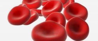
Inflammatory disorders are divided into two large subgroups. The first is infectious. Actually septic diseases. From a common cold to something serious: pneumonia, blood poisoning.
The second is immune changes. When the body's own defenses attack the body's cells. Finding the first process is not that difficult. A routine blood test is sufficient.
To identify the second type of inflammatory process, specific measures are needed. This is a special case that should be considered.
Rheumatoid factor is an immunoglobulin antibody (IgM) that can destroy bacteria and viruses, but its attack is directed at the body's own cells, which are mistaken for foreign cells. A so-called autoimmune reaction occurs.
Rheumatoid factor (RF) is synthesized by the structures of the joint capsules and not only. This is how the body fights an imaginary threat.
There is also a plus here. Using this indicator, it is possible to quickly identify autoimmune processes. A high or low RF level automatically requires a complete diagnosis of the patient's condition.
Treatment is prescribed as needed. It is mainly aimed at combating the root cause. Symptomatic measures also take place.
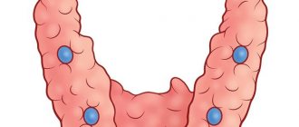
What is rheumatoid factor
Rheumatoid factor is an autoantibody that is produced by the immune system. Autoantibodies cause autoimmune and inflammatory processes by mistakenly attacking the body's own tissues. Rheumatoid factor is synthesized by plasma cells of the synovial membrane of the joint, moves into the blood, where it also affects blood vessels and other synovial membranes.
SCHEME OF INFLAMMATION DEVELOPMENT IN RHEUMATOID SYNOVITIS AND PRODUCTION OF RHEUMATOID FACTOR BY PLASMA CELLS ()
This marker gets its name from its initial discovery in people with rheumatoid arthritis. Since then, it has become apparent that other diseases and health conditions can trigger its production.
Elevated levels of rheumatoid factor have also been found in people with other autoimmune diseases, chronic infections, cancer, liver disease, and parasites. [, ] In particular, increased values of rheumatoid factor are determined in people with: systemic lupus erythematosus, systemic scleroderma, sarcoidosis, dermatomyositis, Sjogren's syndrome, septic endocarditis, infectious mononucleosis, tuberculosis, leprosy, viral hepatitis in the active phase, malaria, leishmaniasis, trypanosomiasis, Waldenström's macroglobulinemia, chronic lymphocytic leukemia, malignant neoplasms, etc.
Rheumatoid factor is a curious autoantibody that attacks other antibodies. It mainly takes the form of an IgM antibody (the largest antibody). Rheumatoid factor can alternatively exist as other types of antibodies (IgG, IgA, IgE, or IgD), but the IgM form is usually the first to appear in the blood and is more closely related to disease activity. [, , ]
SCHEME OF INFLAMMATION AND PRODUCTION OF RHEUMATOID FACTOR IN RHEUMATOID ARTHRITIS
Healthy people can also produce some forms of rheumatoid factors , which is our body's normal defense against bacterial toxins (lipopolysaccharides) and viruses, such as the Epstein-Barr virus . They do not attack the body's own tissues, but instead help fight infections and clear the body of immune cells that are no longer needed. Once the threat ends, their levels naturally drop. [, , ]
But in people with rheumatoid arthritis, this rheumatoid factor remains at elevated levels for longer periods of time. It probably doesn't directly cause the disease, but it does worsen symptoms by increasing inflammation and destroying joints. [, ]
Rheumatoid factor testing is primarily used to help diagnose rheumatoid arthritis and determine how advanced the disease is. Levels of this autoantibody often rise for many years before symptoms appear and may remain high long after treatment. However, rheumatoid factor is not considered the exclusive criterion for determining rheumatoid arthritis; there are a sufficient number of people with rheumatoid arthritis in whom this autoantibody is never detected.
[, , ]
Nephelometric and turbidimetric determination of RF
The methods are based on measuring the intensity of the light flux passing through blood plasma with suspended solid particles. It decreases due to the absorption and scattering of light. Nephelometry and turbidimetry make it possible to assess the “turbidity” of the material under study using a special calibration schedule, determining the amount of IgM-RF in the plasma.
These methods are more informative and accurate than the latex test. They relate to quantitative analyzes and make it possible to reliably determine the concentration of rheumatoid factor in the blood plasma. They are suitable for monitoring the RF level over time. Periodic examinations of the patient make it possible to assess the rate of progression of autoimmune diseases and the effectiveness of the therapy.
ELISA for determining rheumatoid factor IgM, IgG, IgA and IgE
All previous methods are aimed at determining IgM-RF, which makes up 90% of the total pool of pathological immunoglobulins. However, they are not able to detect autoantigens of other classes. The enzyme immunoassay does not have this drawback. Using ELISA, you can detect IgG-RF, IgE-RF and IgA-RF.
An increase in pathological IgG levels usually indicates damage to the vascular endothelium. This is typical for autoimmune diseases accompanied by the development of vasculitis. A high concentration of IgA usually indicates a severe and prognostically unfavorable course of rheumatoid arthritis.
Rheumatoid factor test
Your doctor will routinely order a rheumatoid factor test if you show symptoms of rheumatoid arthritis, such as [, ]:
- Joint pain, tenderness, redness and swelling
- Joint stiffness
- General fatigue and weakness
- Slight increase in temperature
- Dry eyes or mouth
Together with this test, they often also test for antibodies to cyclic citrulline-containing peptide (ACCP), which together makes it possible to very accurately diagnose rheumatoid arthritis.
[]
Determination methods
Methods for determining RF are divided into qualitative and quantitative . The first include the latex test and the classical Waaler-Rose reaction, which is practically no longer used. These tests make it possible to detect with some certainty an increase in rheumatoid factor.
To accurately detect the level of RF, quantitative determination (nephelometric or turbidimetric) is used. An even more advanced test is ELISA - enzyme-linked immunosorbent assay. It allows you to detect the concentration of not only IgM-RF, but also other pathological immunoglobulins. This opens up new diagnostic possibilities and makes the analysis more informative.
Normal rheumatoid factor levels
The normal level for rheumatoid factor varies depending on the reagents used in different laboratories, but generally the normal value should be less than <14 IU/mL (or <20 IU/mL, depending on the laboratory). [,,]
If you believe your rheumatoid factor levels are elevated, tests for certain diseases or conditions may show false readings.
Rheumatoid factor may interfere with the following laboratory tests:
- Malaria (false positive) []
- HIV (false positive) []
- Hepatitis C (false positive) []
- Antibodies to cardiolipin (false positive) []
- Thyroid-stimulating hormone (falsely increased) [, , ]
- Cytokines including IL-1-beta, IL-4, IL-6 and IL-8 (falsely elevated). [ ]
Latex test
The simplest, cheapest and fastest test for which an RF latex reagent containing human IgG is used. Blood serum is taken as the test material. The abnormal immunoglobulins it contains react with the Fc fragments of IgG that are in the reagent.
If the serum contains more than 8 U/ml of rheumatoid factor, a pronounced agglutination reaction occurs (the gluing of normal and pathological immunoglobulins to each other). Visually it can be seen as a positive test. The duration of the study is about 15-20 minutes.
The latex test has its drawbacks. These include low information content and a high frequency of false positive results. Unlike quantitative methods, the latex test does not make it possible to detect the level of RF in the blood plasma.
Causes of elevated rheumatoid factor
Rheumatoid arthritis
Rheumatoid arthritis (RA) is the most common systemic autoimmune disease that causes inflammation and destruction of the joints.
About 70-90% of people with rheumatoid arthritis have rheumatoid factors , which can further stimulate inflammation and damage to body tissue. Patients who test positive for rheumatoid factor show a more intense inflammatory response and worse disease prognosis than those people who do not have these autoantibodies. [,,,]
Higher levels of rheumatoid factor (>50 IU/ml) at the onset of RA disease are also associated with worse prognosis and severe arthritis. [, ]
Reducing this marker may play a positive role in the successful treatment of rheumatoid arthritis . Reducing rheumatoid factor levels has been shown to improve drug response in people with RA. []
NEGATIVE EFFECT OF INFLAMMATORY CYTOKINE IL-6 ON VARIOUS ORGANS IN RHEUMATOID ARTHRITIS ()
Other forms of arthritis
Rheumatoid factor levels may also be elevated in other forms of arthritis, although less likely than in RA.
Psoriatic arthritis occurs in people with psoriasis and rheumatoid factor is found in 15% of patients. []
Juvenile idiopathic arthritis is a form of autoimmune arthritis that occurs in children under 16 years of age. Its cause is still unknown, while rheumatoid factor is found in 5% of children with this disease. []
Other diseases and health conditions
High levels of rheumatoid factor are also found in a variety of diseases and conditions, including [R]
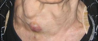
- Sjogren's syndrome is an autoimmune disease that causes dry mouth and eyes (rheumatoid factor is found in 75-95% of cases)
- Primary biliary cirrhosis, an autoimmune disease that destroys the bile ducts (45-70%)
- Mixed connective tissue disease – an autoimmune disease similar to lupus (50-60%)
- Viral infections, including hepatitis, HIV, Epstein-Barr , cytomegalovirus (10-76%)
- Breast cancer (13-47%) []
- Liver cirrhosis, liver fibrosis (25%)
- Lupus erythematosus is an autoimmune disease that damages connective tissue (15-35%)
- Sarcoidosis is an inflammatory disease that damages the lungs and lymph glands (5-30%)
- Parasitic infections such as malaria and toxoplasmosis (10-18%)
- Bacterial infections, including chlamydia, syphilis and tuberculosis (8-15%)
Lifestyle factors
Smoking
Smoking is the strongest environmental risk factor for the development of rheumatoid arthritis. [,]
An observational study of 296 people found that 88% of smokers tested positive for rheumatoid factor within 10 years. There were also more smokers in the rheumatoid-positive group than in the rheumatoid-negative group. []
The risk of elevated rheumatoid factor levels was 4 times higher in smokers than in people who had never smoked (observational study of 7,100 people). Another study of 100 patients with rheumatoid arthritis found a 2-fold increase in rheumatoid factor in former and current smokers compared to those who had never smoked. [, ]
Smoking approximately 1 pack (20 cigarettes) per day for 25 years leads to a 3-fold increase in the risk of high rheumatoid factor (study of 336 patients with rheumatoid arthritis). Moreover, rheumatoid factor levels were directly related to the number of years of smoking. The longer a person smoked, the higher the level of rheumatoid factor (2 studies with a total of 673 people with rheumatoid arthritis). [ , , ]
A lot of coffee
The number of cups of coffee consumed daily was directly associated with the risk of developing rheumatoid arthritis (RA) and positive rheumatoid factor testing in an observational study of 19,000 people. who drank 4 or more cups of coffee per day had more than twice the risk of developing rheumatoid arthritis and increased rheumatoid factor levels compared to people who drank less coffee. []
Aging
Rheumatoid factors are found at elevated levels more often in older adults (10-25% of all older participants) than in younger adults (5% of younger participants), as aging can cause a gradual imbalance of the immune system. [, , ]
OBESITY INCREASES THE RISK OF RHEUMATOID ARTHRITIS, INCREASES INFLAMMATION AND SEVERITY OF THE DISEASE ()
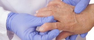
When is treatment required?
Before treating any joint or autoimmune disease, you need to make sure it is present. Detection of a high content of rheumatic factor in the blood is not a basis for making a diagnosis. We can talk about the disease only if there are characteristic symptoms and the results of other, more reliable tests. Treatment should begin only after confirmation of the diagnosis. All medications must be prescribed by doctors.
To combat collagenosis, glucocorticosteroids and cytostatics are usually used. These drugs inhibit the activity of the immune system and inhibit the synthesis of autoantibodies. For severe rheumatoid arthritis, the use of biological agents (Rituximab, Humira, Embrel, Remicade) is very effective. To combat infectious diseases, a course of antibacterial, antiviral or antiparasitic therapy is required.
People with Sjögren's disease require symptomatic treatment for dry eye syndrome. For this purpose, they are prescribed artificial tears. With concomitant damage to the thyroid gland, the patient may need to take Eutirox, a synthetic analogue of its hormones.
Health effects of high rheumatoid factor levels
Risk of rheumatoid arthritis
One observational study that followed 9,700 healthy people for 30 years found that slightly elevated levels of rheumatoid factor (25-50 IU/ml) were associated with an almost 4-fold increase in the risk of developing rheumatoid arthritis (RA). Very high levels (>100 IU/mL) were associated with a 26-fold increased risk compared with normal levels (up to 25 IU/mL). []
Risk of deep vein thrombosis
Rheumatoid factor levels above 110 IU/mL were associated with a 3-fold increased risk of blood clots in the deep veins (deep vein thrombosis) in an observational study of 670 people. []
Risk of atherosclerosis
One observational study of 6,500 people found that rheumatoid factor levels exceeding 15 IU/mL resulted in an increased risk of atherosclerosis in African-American women. []
PROLONGED INFLAMMATION IN RHEUMATOID ARTHRITIS LEADS TO: DEVELOPMENT OF METABOLIC SYNDROME (INCREASED INSULIN RESISTANCE), OSTEOPOROSIS, CARDIOVASCULAR DISEASES ()
Poor recovery after stroke
Rheumatoid factor levels within the normal range (above 7.6 IU/mL) were associated with a 79% increased risk of brain dysfunction compared with barely detectable levels (below 1.1 IU/mL) in a study of 582 patients with stroke. []
Risk of Cardiovascular Diseases
Men with rheumatoid factor levels above 6 IU/mL had a 2.9-fold increased risk of developing heart disease compared with men with levels below 6 IU/mL in a study of 1,100 healthy people. This association was not observed in women. []
Death from heart disease, cancer or any other cause
Negative rheumatoid factor test results were associated with a lower risk of death. In one observational study of 12 patients, positive (positive) rheumatoid factor demonstrated a 31% increased risk of death from any cause. Detectable levels of RF were also associated with a 45% increased risk of death from heart disease. []
In another study of nearly 300,000 people, RF levels greater than 20 IU/mL were associated with a 50% increased risk of death from any cause and a 56% increased risk of death from cancer . []
Another observational study (25 years) of 2,900 patients with rheumatoid arthritis showed an increase in these effects. Those people who tested positive for rheumatoid factor had almost twice the risk of premature death than those who tested negative for rheumatoid factor. []
TEMPORARY RELATIONSHIP BETWEEN THE PRODUCTION OF THE HORMONE CORTISOL AND INCREASE OR DECREASE IN ACTIVITY OF RHEUMATOID ARTHRITIS ()
CRP for cancer
In oncology, CRP is a nonspecific indicator. It does not make it possible to determine where exactly the cancer is developing. It increases due to inflammation caused by the growth of a malignant tumor. To determine the location of cancer, you need to take specific tumor markers, do an ultrasound, computed tomography or magnetic resonance imaging.
PSA is determined when diagnosing recurrent ovarian and skin cancer. In colon cancer, protein is a prognostic criterion. The higher the indicator, the lower the survival rate of patients.
Ways to reduce rheumatoid factor
Quit smoking
Smoking is directly associated with an increased risk of elevated rheumatoid factors and the development of rheumatoid arthritis . [R, R, , , , , , ]
If you are a smoker and have high levels of rheumatoid factor (or a family history of autoimmune diseases such as rheumatoid arthritis), it is imperative that you stop smoking, which can help reduce your risk of poor health and the development of autoimmune diseases.
Reduce coffee consumption
If you're an avid coffee drinker, gradually reducing the amount of coffee you drink each day, or avoiding coffee altogether, can help lower your rheumatoid factor levels and reduce your risk of rheumatoid arthritis.
Vegetarian diet
A study of 53 people found that following a vegetarian diet for one year resulted in a greater reduction in rheumatoid factor levels than an omnivorous diet. []
Yoga
One week of yoga practice led to 8% reduction in rheumatoid factor levels in a study of 64 patients with rheumatoid arthritis. []
Fish oil and olive oil
A combination of fish oil and olive oil for 24 weeks reduced rheumatoid factor levels by 30% in a study of 43 people with rheumatoid arthritis. [ ]
Vitamin E
In a study of 50 people who took 600 mg of vitamin E (dl-α-tocopheryl acetate) per day for 6 weeks, rheumatoid factor levels decreased by 44%. []
Vitamin K1
Taking 10 mg per day of vitamin K1 for 8 weeks reduced rheumatoid factor levels by 16% in a study of 64 patients with rheumatoid arthritis. []
Curcumin
In a study of 12 patients with rheumatoid arthritis, taking 1000 mg of curcumin per day reduced rheumatoid factor levels over 90 days. If you choose to supplement with curcumin, then purchase bioactive forms that have better absorption in the intestines. []
Ashwagandha, ginger, boswellia, turmeric
An Ayurvedic combination of ashwagandha, ginger, boswellia and turmeric, when used by 165 patients with rheumatoid arthritis for 16 weeks, helped reduce rheumatoid factor levels along with reduced joint swelling. []
Andrographis
Andrographis is a popular herbal supplement often used in Southeast Asia to reduce pain, inflammation and fever in Traditional Chinese Medicine and Ayurveda. In a study of 60 people with rheumatoid arthritis, taking andrographis for 14 weeks reduced rheumatoid factor levels. []
Annual wormwood (Artemísia annua)
Artemisia annua extract reduced rheumatoid factor levels in 159 people with rheumatoid arthritis (clinical study). []
Thunder God Vine (Tripterygium wilfordii)
Tripterygium wilfordii is a plant used in traditional Chinese medicine as a remedy for various autoimmune and inflammatory diseases.
In a meta-analysis of 6 studies and a total of 643 participants in people with rheumatoid arthritis, this plant reduced rheumatoid factor levels. []
Some aqueous extracts of this plant can have a negative effect on intestinal function, stimulate the appearance of rashes, and reduce the number of white blood cells. Almost all of these negative effects can be avoided by using safer alcohol extracts and taking them in the correct dosage. [, ]
Pine pollen
Pine pollen extracts reduce rheumatoid factor levels and other markers of inflammation in mice with arthritis. []
Resveratrol
In arthritic rats, resveratrol reduced rheumatoid factor levels by 63%. []
Alginic acid (brown seaweed)
Alginic acid, found in brown, red and green seaweeds, reduced rheumatoid factor levels in rats with rheumatoid arthritis. []
Arisema rhizomatum
Arizema rhizome is a traditional Chinese medicine herb that is used to reduce pain and inflammation. It markedly reduced rheumatoid factor levels in a study of arthritic rats. []
SRB norm
In the blood of a healthy person, the level of CRP is very low or this protein is completely absent (in a laboratory test, but this does not mean that it is not there at all - the test simply does not detect tiny amounts).
The following limits of values are accepted as the norm, and they do not depend on age and gender: for children, men and women it is one - up to 5 mg/l, the only exception is newborn children - they are allowed to have up to 15 mg/l of this acute-phase protein (as evidenced by reference literature). However, the situation changes if sepsis is suspected: neonatologists begin urgent measures (antibiotic therapy) when the child’s CRP increases to 12 mg/l, while doctors note that a bacterial infection in the first days of life may not cause a sharp increase in this protein.
A laboratory test is prescribed to detect C-Reactives protein in the case of many pathological conditions accompanied by inflammation, the cause of which is infection or destruction of the normal structure (destruction) of tissues:
- Acute period of various inflammatory processes;
- Activation of chronic inflammatory diseases;
- Infections of viral and bacterial origin;
- Allergic reactions of the body;
- Active phase of rheumatism;
- Myocardial infarction.
In order to better understand the diagnostic value of this analysis, it is necessary to understand what acute phase proteins are, learn about the reasons for their appearance in the patient’s blood, and consider in more detail the mechanism of immunological reactions during an acute inflammatory process. Which is what we will try to do in the next section.
What to do and how to treat elevated C-reactive protein?
An increased concentration of CRP, confirmed by a biochemical blood test, is not an accurate confirmation of a specific disease. This is an indicator of the development of a possible pathology. What it may be connected with can only be determined based on additional research.
It is noteworthy that if the therapy is chosen correctly, the level of C-reactive protein quickly decreases and returns to normal. For example, with the correct use of antibacterial drugs, a positive result is noted by a decrease in the level of CRP within 24 hours. If there are no obvious signs of bacterial or viral infection, but the analysis shows an increased concentration of CRP in the blood, then a consultation with an oncologist is required.
In order for any prescribed therapy to be effective, you should follow the rules of a healthy diet and do not forget about moderate physical activity. In addition, you need to try to eradicate existing bad habits. Such standard rules will contribute to rapid recovery and preservation of health for many years.
Antistreptolysin-O (ASLO, ASO)
Antisteptolysin-O are antibodies (AT) directed to streptolysin (the so-called antigen of beta-hemolytic streptococcus). The norm for adults is up to 200 U/l; for children under 14 years old – up to 150 U/l.
To determine antistreptolysin-O and its titers (the kit includes a separate titration kit), there is also a latex test - the study is quick and not labor-intensive, but some doctors prefer to conduct a blood test using the turbidimetric method, as they have more confidence in it. But this method (turbidimetric) already requires the use of more complex equipment.