Hemorrhagic syndrome is a complex symptom complex caused by dysfunction of the hemostatic system, which normally preserves the liquid state of the blood, maintains the structural integrity of the vascular wall and ensures its rapid thrombosis when damaged. The syndrome is a clinical manifestation of various diseases and is characterized by a variety of etiopathogenetic factors. In patients with impaired hemostasis, bleeding increases, external and internal bleeding occurs. The most common type of the latter is hemorrhage - the release of blood from a vessel and its accumulation in tissues, organs and cavities. The syndrome in acute and chronic forms has significant differences in terms of diagnostic criteria, principles of medical care and prognosis for recovery.
Persons of any age are susceptible to the development of hemorrhagic syndrome - both newborns and elderly people. The syndrome affects women much more often than men. The reasons for its development are internal diseases and external influences. Blood flows from the blood vessel through the damaged area. In patients, hemorrhages appear on the skin, mucous membranes begin to bleed, and internal bleeding occurs. The symptoms of the anomaly are determined by the location of the existing defect.
The acute form requires emergency treatment, and the chronic form requires an integrated approach. The main goal of therapeutic measures is to improve the blood clotting process. When patients lose a lot of blood, immediate medical attention is necessary.
Development mechanism
The formation of a pathological process is based on a group of provoking factors. Generally speaking, the following points can be mentioned:
- Genetic abnormalities. They occur relatively often. They account for almost 25% of the total clinical situations. The classic version is hemophilia: an incurable disease in which normal blood clotting is disrupted.
It makes sense to examine the patient for genetic abnormalities, although this is not always possible due to the complexity of diagnosis. Treatment in this case comes down to correction of symptoms; etiotropic effects are impossible for obvious reasons.
- Coagulation factor disorders. There is a whole group of pathologies that are typically characterized by a disruption in the production of special substances responsible for coagulation. These include von Willebrand's disease, disseminated intravascular coagulation syndrome and other similar disorders, including the above-mentioned hemophilia.
Attention:
Diseases of this kind are difficult to correct or cannot be corrected at all. Symptoms are also difficult to relieve.
- Pathologies and inferiority of the vessels themselves, capillaries and larger structures. Increased permeability, fragility. Occurs as a result of congenital hereditary anomalies or acquired problems. For example, with a lack of vitamins B and C, the normal structure of blood vessels is disrupted, and massive bleeding occurs, including fatal ones.
- Other factors that are brought to life by the patient himself or the environment. As a rule, they are transient and can be easily corrected on their own. If there is an understanding of what was the immediate cause of the problem.
Mixed variants of pathogenesis are also often found. In this case, you need to influence all provoking factors in parallel.
Violations of normal coagulation or problems with the structure of blood vessels, their integrity, strength, elasticity, physical properties lead to a pathological disorder of hemostasis.
The blood does not stop on its own, or the capillaries or other structures begin to collapse in large quantities, causing the formation of a specific rash and the release of liquid connective tissue into the joints. Severe disability, emergency conditions, and death are possible.
The pathogenesis of the hemorrhagic process is multiple, caused by congenital or acquired disorders of coagulation and vascular structure.
If you detect at least some symptoms reminiscent of hemorrhagic syndrome, you should immediately consult a doctor.
Etiopathogenesis
Hemorrhagic syndrome is the result of disruption of the hemostasis system. Usually the walls of blood vessels are affected, the structure and number of platelets change, and coagulation processes are disrupted.
According to the etiopathogenetic classification, there are two main forms of the syndrome:
- The hereditary or primary form is genetically determined: the patient has a defective gene that encodes the functioning of the entire hemostatic system or its individual elements - platelet cells, plasma factors and vascular endothelium. Hereditary forms include thrombocytopathies, hemophilia, von Willebrand-Diana disease.
- The acquired or secondary form is a consequence of inflammation of autoimmune origin that affects the vascular wall, as well as various injuries, metabolic disorders or severe drug intoxication. Patients are most often diagnosed with secondary thrombocytopenia and thrombocytopathy, DIC syndrome. This also includes medicinal and psychogenic forms. The first is associated with taking drugs that suppress the process of platelet aggregation and coagulation - the sticking together of small particles into larger ones. The second form is caused by patients themselves due to mental disorders.
There are a number of diseases that manifest signs of the syndrome. These include:
- hepatitis,
- oncopathology,
- acute infections of viral etiology,
- cirrhosis of the liver,
- leukemia,
- vasculitis,
- hemorrhoidal syndrome.
Frequent blood transfusions, traumatic injury, and shock can provoke the development of pathology.
In newborns, the disease develops as a result of the following reasons:
- use by a pregnant woman of drugs from the group of salicylates, phenobarbital, antibiotics, anti-tuberculosis drugs,
- pathological pregnancy,
- oxygen starvation of the fetus in the womb,
- prematurity,
- neoplastic damage to connective tissue fibers,
- lack of vitamin K,
- malabsorption syndrome,
- the mother has high-risk diseases.
Hemorrhagic syndrome in a newborn can manifest itself clinically on the first day of life, but more often symptoms appear on days 5-7.
Classification
The division is carried out according to a group of bases. The first and most important thing that is used in clinical practice is the dynamics of the disorder.
Accordingly they are called:
- Angiomatous form. Accompanied by severe bleeding at the site of the lesion. A typical feature of this type of disease is focal destruction of blood vessels, without involvement of neighboring areas.
However, the nature of the leakage of liquid connective tissue makes angiomatous hemorrhagic syndrome deadly, since massive internal bleeding is likely (if a large vessel is affected).
- Hematoma variant. Reminiscent of the previous one, however, much more formidable in terms of prognosis. Develops against the background of DIC syndrome, in the hypocoagulation phase, also hemophilia, in the acute period. Symptoms include the development of massive, profuse hemorrhages in the tissue.
Joints, organs, and brain suffer. Without systematic treatment aimed at preventing such a scenario, a person’s death from complications is likely. It is the matter of time.
- Vasculitic purpuric form. Accompanied by the formation of small hemorrhages on the skin of a purple or dark red hue. The petechial rash itself, passing on its own, leaves pigmented areas and cosmetic defects.
This form does not pose any great danger. However, it develops as a result of infectious inflammatory processes affecting blood vessels. Therefore, it is quite possible for hemorrhage to transform into disseminated intravascular coagulation syndrome; this is already a deadly disorder.
- Petechial type. The classic form of the pathological process. Accompanied by massive rashes in the form of small circles 2-5 mm in diameter. Dark purple or deep red.
Recovery is not possible because a number of clotting factors are involved. As part of symptomatic correction, replacement drugs are used.
This form of pathology behaves unpredictably, therefore it is extremely difficult to predict its course.
- Mixed type. For him, the development of a group of manifestations is pathognomonic. On the other hand, there is also a feature characteristic only of this form. Despite hemorrhages in organs, tissues, and the formation of a rash, the joints are practically not involved.
There are also atypical, rarely encountered species. For example, thrombohemorrhagic syndrome, which is characterized by alternating episodes of hypocoagulation and active, excessive production of coagulation factors with the formation of clots blocking blood vessels (thrombi).
Another way of classification is by the origin of the disease.
- Congenital appearance. Occurs from the moment a child is born. It has hereditary, genetic roots. It is practically not subject to restoration, not counting the most successful cases.
- Acquired form. For some pathologies: von Willebrand, disseminated intravascular coagulation and others. The prospects for therapy are different; anything can be said for sure after assessing the specific clinical situation.
According to the nature of the flow:
- Sharp look. A critical hemorrhagic condition occurs with severe hemorrhages, the formation of a skin rash, symptoms of general intoxication of the body, weakness, muscle and joint pain.
- Chronic form. It proceeds sluggishly, gives a minimum of clinical manifestations, but at any moment it can go into a critical phase. It is poorly diagnosed; as a rule, the patient does not see a doctor until the last moment. Possible accidental detection during a routine examination.
More detailed classifications exist, but they are used less frequently in clinical practice or are not considered generally accepted in medicine.
Prevention
Prevention of hemorrhagic syndrome begins in the maternity hospital. Vitamin K is intended to prevent hemorrhages in premature newborns, administered to babies for the purpose of prevention in a minimal dose (1 mg) immediately after birth.
In order to prevent hemophilia in families where cases of the disease have occurred, future parents should visit a geneticist to calculate the risk of having a sick child.
If there is increased bleeding or taking blood-thinning drugs, the patient is obliged to report these circumstances to dentists, surgeons and other specialists whose actions may lead to bleeding.
Symptoms
The clinic depends on the specific form of hemorrhagic syndrome. It must be assessed accordingly.
Angiomatous variety
- Pain at the site of bleeding. Insignificant in character, weak. Aching, pulling. With a massive process, the phenomenon can be much stronger. Unstable in terms of current. They become pronounced when moving, changing body position, or physical activity.
- An outpouring of fluid connective tissue in a clearly localized area. This is a characteristic distinguishing feature of the angiomatous form from others. There is a delimited area of damage. However, it is not always possible to detect it by eye. Diagnosis required. The symptom is not felt subjectively.
- Swelling of the affected area. With a significant amount of blood released, an enlargement of the localized area occurs. How strong depends on the nature and intensity of the process.
- Weakness, drowsiness, lethargy.
With prolonged course, manifestations of anemia are possible. Skin problems, brittle hair, shortness of breath, heart disorders, exercise intolerance. The result of iron loss.
Hematoma variant
Much more dangerous. Can cause rapid death without treatment.
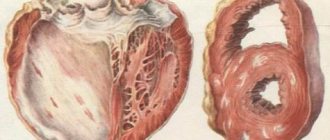
- Massive hemorrhages in the joints (hemarthrosis). The calling card of this type of violation. It is accompanied by a significant increase in the size of the cavity, compression of the cartilage and bursa, and problems with providing local structures with nutrients.
Motor activity disappears, as does the ability to use the affected limb. As a rule, hematoma hemorrhage involves large joints: knees, elbows, hips. Variations are possible.
- Severe pain at the site of the lesion. It intensifies when palpation is attempted. Movement, changing the position of the arm or leg is impossible due to intense discomfort and swelling. A puncture is required, drainage of the joint cavity and restoration of its anatomical shape and functions.
- Formation of large hematomas. On large areas of fabric. They appear as purple or violet, bluish, red areas under the skin. They protrude strongly above the surface of the dermis and change the external properties of the body. Urgent medical attention is required.
- Weakness, drowsiness. As a result of a decrease in hemoglobin concentration. With the development of bleeding in the area of internal organs, focal disorders are possible. For example, problems in the digestive tract, neurological deficits.
- Necrosis with long-term preservation of blood volume in tissues. As a result of ischemia (malnutrition) and compression of surrounding structures. The most dangerous complication. Possible regardless of the location of the process. In particular, with a cerebral hemorrhage (for example, subarachnoid), symptoms reminiscent of a major stroke cannot be avoided. Provided that the elimination of the consequences is not started in a timely manner.
Vasculitic purpuric form
- Massive rashes all over the body. They look like small spots of violet, lilac shades, possibly turning blue. They rarely exceed 1 cm in diameter. They are completely painless, not accompanied by itching, burning, or other discomfort, which distinguishes them from rashes due to dermatitis and skin diseases themselves. They pass independently without any deviations or special events.
- Weakness, weakness.
- Massive bleeding is possible: uterine, intestinal, pulmonary. Depends on the reason that brought the pathological process to life.
Dangerous complications are typical for this form. The likelihood of such happening is difficult to predict in advance. It all depends on the duration of the process, the main reason, and other factors.
Petechial type
This variety is characterized by the development of a massive rash throughout the body, small red or scarlet spots. Up to 3 mm. Also, in addition to the dermal layers, they affect the mucous membranes. They can form, including on internal organs.
Danger to health and life is relatively rare. However, everything depends on the primary disease.
Mixed view
It manifests itself as hemorrhages in the tissue, as well as cutaneous hemorrhagic syndrome, the formation of spots, petechiae, and purpura. As stated, the development of articular lesions is atypical.
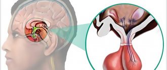
Coma due to hemorrhagic stroke
Approximately 90% of patients with GI in a state of stupor or coma die in the first five days, despite intensive therapy
Disorders of consciousness are characteristic of many pathologies, manifested by inhibition of the functions of the reticular formation of the brain.
Brain dysfunctions develop under the influence of:
- Endo- and exotoxins – derivatives of the end products of metabolism;
- Oxygen and energy starvation of the brain;
- Metabolic disorders in brain structures;
- Expansion of the volume of brain matter.
The most important factors in the development of coma are acidosis, cerebral edema, increased intracranial pressure, and impaired microcirculation of brain fluids and blood.
The state of coma affects the functioning of the respiratory system, excretion (kidneys) and digestion (liver, intestines).
It is impossible to recover from a coma at home, and it is very difficult even in intensive care conditions.
The clinical definition of coma is carried out using the GCS (Glasgow Coma Scale), and some other methods that are important for clinicians are used. There are precoma and four stages of coma. The easiest is the first, and the hopeless state of the patient corresponds to the fourth stage of coma.
Causes
If we summarize the possible provoking factors, the following picture emerges:
- Disorders of coagulation processes. Congenital (much more often) and acquired. Hemophilia, other pathological conditions.
- Changes in the concentration of formed cells or their inferiority and inability to perform their own functions (thrombocytopenia - pathies). They occur quite often and can be transient, temporary phenomena.
- Vasculitis, inflammatory processes of blood vessels. Infectious or autoimmune forms.
- Disorders of the liver and kidneys. Especially cirrhosis, hepatitis, failure. In this case, hemorrhagic syndrome is considered secondary.
- Tumors of various localizations. Regardless of the type. Typically malignant.
- Genetic abnormalities.
- Mixed diseases such as von Willebrand pathology, DIC syndrome. They have a multiple nature and are accompanied by unstable symptoms from both coagulation factors and the structure of blood vessels.
What are hemorrhages
In medicine, spontaneous bleeding from blood vessels in any part of the body is called hemorrhage. This pathological syndrome manifests itself in patients in response to external influences or in the presence of internal diseases. Hemorrhagic disease occurs due to damage to the integrity of the walls of blood vessels, a decrease in the number of platelets, and a violation of coagulation hemostasis. In this case, blood flows beyond the boundaries of the blood vessel through the damaged area. The types of abnormalities depend on which part of the body they appear in.
Hemorrhagic syndrome is typical for what diseases?
Among the forms of hemorrhagic diseases, hereditary and acquired disorders of hemostasis are distinguished. The latter are associated with multifactorial disorders of the blood coagulation system (for example, acute disseminated intravascular coagulation syndrome), damage to blood vessels of dysmetabolic, immune, toxic-infectious, immune complex origin, abnormalities of adhesive proteins in blood plasma, damage to platelets and megakaryocytes. Hereditary hemorrhagic diseases are caused by:
- pathologies of plasma factors of the blood coagulation system;
- hereditary disorder of hemostasis;
- genetic structural changes in the vascular wall.
Diagnostics
The examination is carried out within the walls of the hospital, especially in acute cases. The main specialist is a hematologist.
Typical activities:
- Oral interview with the patient. It is necessary to identify and record all complaints for their further assessment and analysis.
- Anamnesis collection. As part of the initial examination, it is necessary to determine the likely origin of the anomalous process. The technique allows you to detect the nature of hemorrhagic syndrome.
- General blood test, biochemistry. Provides information on the composition and rheological properties of the fabric.
- A coagulogram is mandatory. Coagulation rate study. Provides an opportunity to recognize the existence of a problem.
- Ultrasound of internal organs, joints. As part of assessing the condition of anatomical structures, excluding latent hemorrhages.
It is possible to conduct genetic tests and analyzes as needed. If the doctor deems it necessary.
The list of events is incomplete, however, they are held most frequently.
Diagnostic measures
Diagnosis of the syndrome is aimed at establishing its cause for its further elimination during treatment. It lies in the correct assessment of clinical signs and results of laboratory and instrumental studies. Specialists begin diagnosis by carefully collecting anamnesis, listening to the patient’s complaints and carefully examining his skin.
- During the interview, doctors are interested in the presence and nature of bleeding, and during the examination they examine the rash on the skin and hematomas near the joints.
- Taking an anamnesis is necessary to determine the primary or secondary nature of the disease, the presence of concomitant pathologies, and the time of onset of the main symptoms.
- The coagulogram determines coagulation factors, platelet and coagulation components of hemostasis, the level of fibrinogen and prothrombin.
- In a general blood test, the number of platelets is counted, signs of inflammation are detected, and in an immunogram, the level of immunoglobulins of each type is detected.
- Ultrasound examination, sternal puncture and histological analysis of bone marrow biopsy are carried out according to indications.
If a patient is admitted to a hospital with an acute form of pathology, he requires emergency care - stopping bleeding and restoring hemostasis. Such patients are diagnosed after emergency treatment, when their condition becomes stable.
Treatment
Therapy is designed to solve two problems: combating the root cause, correcting symptoms.
Symptomatic supervision. The key direction is implemented through procedures and the use of drugs:
- Glucocorticoids. Stop inflammatory processes, if present. Excellent for the treatment of a number of forms of thrombocytopathy.
- Immunosuppressants. As part of eliminating complex autoimmune conditions. In particular, vasculitis and others.
- Saline solutions. To restore water balance and electrolyte concentration.
- Heparins as needed. For blood thinning during thrombotic phases of DIC syndrome.
- Iron preparations. To eliminate anemia.
- Vitamin complexes. Ascorutin, medicines based on C, B.
If necessary, donor coagulation factor preparations are used, plasma and red blood cells are transfused.
Surgical treatment is performed if necessary. Drainage of hematomas, pumping out blood. These are symptomatic measures. Manifestations are eliminated with the help of replacement agents, also iron-based products. There are many options.
In the future, systematic examinations by a hematologist are indicated, at least 2 times a year. Some disorders do not respond to effective treatment. For example, hemophilia and a number of others.
Treatment process
To cope with hemorrhagic syndrome, it is necessary to determine its cause. Treatment tactics for the syndrome are chosen depending on its variant, stage, severity of the process and the presence of concomitant disorders.
All patients are hospitalized in a medical facility, the bleeding is stopped, the deficiency of vitamin K is compensated, and then they begin drug treatment, the purpose of which is to restore the full functioning of the blood coagulation system. The attending physician prescribes medications and selects therapeutic procedures individually for each patient. During medical procedures, continuous monitoring of pressure, temperature, pulse and respiration is necessary.
Treatment regimen for hemophilia :
- Blood transfusion therapy - blood transfusion directly from a donor, administration of fresh frozen plasma;
- Direct-acting coagulants are chromatographically purified lyophilized preparations of human blood plasma containing coagulation factor VIII and complex factor IX, as well as a recombinant preparation of factor VII;
- Hemostatic drugs - direct hemostatics “Fibrinogen”, “Thrombin”, indirect coagulants “Menadion”;
- Injection of corticosteroids "Kenalog" or "Diprospan" into the joint cavity after aspiration of blood;
- During remission - exercise therapy, magnetic therapy, electrophoresis and other absorbable physiotherapeutic procedures,
- In case of complications - surgical intervention.
General treatment regimen for coagulopathy :
- Antihemorrhagic agents - “Vikasol”, “Ditsinon”, “Kontriven”;
- Fibrinolysis inhibitors – “Aprotinin”, “Aminocaproic acid”, “Tranexamic acid”;
- Stimulator of platelet adhesion and aggregation – “Etamzilat”;
- Infusion of plasma, platelet or red blood cells, blood substitutes, colloid and crystalloid solutions;
- "Somatotropic hormone."
Treatment of DIC syndrome :
- "Heparin" in the hypercoagulation stage,
- "Kontrikal" in the hypocoagulation stage.
For thrombocytopenic purpura, patients are prescribed:
- Hormone therapy with corticosteroids - Prednisolone,
- Chemotherapy with cytostatics - Cyclosporine,
- Plasmapheresis,
- Splenectomy.
Hemorrhagic telangiectasias are treated conservatively using minimally invasive techniques - sclerotherapy or ozone therapy, as well as surgically - using laser correction, electrocoagulation, cryodestruction with liquid nitrogen or radio wave surgery.
All patients, regardless of the variant of the syndrome and its root cause, are prescribed iron-containing drugs - Tardiferon, Heferol, Ferronate.
Symptoms
The pathology clinic includes signs combined into several syndromes:
- Skin syndrome is characterized by the appearance on the skin of small pinpoint red blood rashes - petechiae, large bruises - ecchymoses and hematomas.
- Internal and external bleeding - nasal, gingival, esophageal, uterine, gastrointestinal.
- Hemorrhages into the joint cavity and intermuscular space, partially or completely immobilizing the patient.
- Pain syndrome of varying severity.
- Anemic syndrome is the result of blood loss.
There are various clinical variants of the pathology: hematoma, petechial-bruise, mixed and others. They all have different symptoms and etiology.
- The hematoma variant is very difficult for patients to tolerate. It occurs in hemophilia and is characterized by profuse bleeding into the muscles and joints. This happens spontaneously or under the influence of even a minor traumatic factor. The pelvic, knee, shoulder and elbow joints are most affected. The main manifestations of the disease are: severe pain, swelling of the periarticular tissues, deformation and dysfunction of the joint, limitation of the patient’s physical activity. During diagnosis, a positive symptom of fluctuation is determined, indicating the presence of fluid in a closed joint cavity - blood. Hemarthrosis is always accompanied by the appearance of large hematomas on the skin. The consequences of the hematoma form are chronic synovitis, destruction of cartilage and bones, deforming arthrosis.
- The petechial-spotted variant develops when the structure and number of platelet cells changes. Deformed platelets or their deficiency are the causes of bleeding disorders. This type of disorder is manifested by small bright red spots that appear on any part of the body and do not disappear with pressure. Even minor trauma to the patient leads to intradermal hemorrhages and pinpoint hemorrhages.
- The mixed version is characterized by hemorrhages in the intermuscular space and inside the skin with the appearance of large hematomas and pinpoint petechiae. In this case, there are no intra-articular hemorrhages and signs of hemarthrosis. Bruises are usually extensive, painful and dense.
- The vasculitic purpuric variant is accompanied by the appearance of a bright red or bluish rash on the pigmented skin of the legs. Its elements are small in size and have a dense consistency. They are surrounded by a pigmented rim, rise above the skin, gradually die off and become covered with crusts. This form is caused by autoimmune or infectious inflammation of the microvascular endothelium - capillaries, arterioles and venules.
- The angiomatous variant is caused by damage to the vascular wall and has specific symptoms. Distinctive features of this type are the recurring, persistent nature of bleeding, vascular dysplasia, and the absence of subcutaneous hemorrhages. Most often, patients complain of bleeding from the nose, which in this case is considered life-threatening. Stomach, intestinal, and pulmonary bleeding are possible.
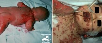
- The edematous-hemorrhagic variant of the syndrome develops in newborns who have experienced hypoxia in utero and is characterized by changes in the lung tissue. Newly born children show signs of respiratory failure in the form of bloody foam from the respiratory tract. Sick children die immediately after birth.
The acquired form of the syndrome is a manifestation of various diseases. That is why its clinical picture includes nonspecific symptoms characteristic of the primary pathological process.
In newborns the syndrome manifests itself:
- hematuria,
- discharge of blood from the vagina,
- constant appearance of blood from the umbilical wound,
- frequent nosebleeds,
- bleeding in the brain.
Clinical guidelines
If the disorder is acquired, a nutritious diet is recommended. The diet should contain large quantities of foods containing a lot of protein and vitamin K.
As aids you can use:
- green apples;
- berries (cherries, raspberries, currants, red grapes, rowan).
In severe forms of the disease, it is necessary to be under constant medical supervision and follow all recommendations of a specialist. The patient should avoid activities that could cause injury. Systematic gymnastic exercises and physiotherapeutic procedures - electrophoresis, magnetic therapy - are effective.
Objective examination
Examination of the skin and mucous membranes.
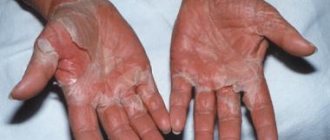
Pinpoint hemorrhages in the skin and mucous membranes (petechiae, ecchymoses) indicate a violation of platelet formation or changes in the vascular wall;
Petechiae
– a type of hemorrhagic spots that appear on the skin and mucous membranes as a result of minute capillary hemorrhages. They appear in varying numbers, usually symmetrically, on any part of the skin, mainly on the extensor surface of the lower extremities (legs, feet), as well as on the mucous membrane of the oral cavity; They have a round shape, a diameter of 1-2 mm, and do not disappear when the skin is pressed or stretched. Their color changes from crimson-red at first to bluish, and then greenish and brown-yellow; are one of the characteristic signs of hemorrhagic diathesis.
Ecchymoses
- hemorrhagic spots are larger in size than petechiae, usually of irregular shape.
Hematomas
- asymmetrically located extensive hemorrhages on the skin indicate a coagulation disorder.
The types and severity of bleeding established during the examination facilitate the diagnostic search.
There are 5 types of bleeding:
1) hematoma;
2) petechial-spotted (microcirculatory, bruise);
3) mixed microcirculatory-hematoma (bruise-hematoma);
4) vasculitic purpuric;
5) angiomatous.
For hematoma type
massive, deep, intense and very painful hemorrhages in large joints, muscles, subcutaneous and retroperitoneal tissue predominate. They cause tissue dissection and destruction, the development of deforming arthrosis, contractures, pathological fractures, bone pseudotumors, and muscle atrophy. Profuse spontaneous, post-traumatic and postoperative bleeding are observed. Characteristic almost exclusively of hereditary coagulopathies, hemophilia A and B.
Petechial spotted
(microcirculatory, bruise)
type
is characterized by painful, non-strained, superficial hemorrhages in the skin and mucous membranes, petechiae, bruises, gingival, nasal and uterine bleeding that do not compress the surrounding tissues. Bleeding occurs with minor trauma to microvessels: when measuring blood pressure, in areas of palpation, when rubbing the skin with a hand, etc. For this type of bleeding, the formation of hematomas is uncharacteristic; muscles, joints and other parts of the musculoskeletal system are intact. Bleeding during abdominal surgical interventions is rare and does not tend to recur. This type of bleeding is observed in thrombocytopenia and thrombocytopathy.
Mixed microcirculatory-hematoma (bruise-hematoma) type
bleeding is characterized not just by a combination of signs of the two variants of hemorrhagic syndrome listed above, but also by a number of qualitative features inherent only to it: petechial-spotted bleeding predominates in the clinical picture; hematomas are few in number, but reach large sizes and are located mainly in the subcutaneous or retroperitoneal tissue; hemorrhages in the joints are rare and do not lead to the development of deforming arthrosis and muscle atrophy; hematomas, depending on the location, can imitate the picture of an acute abdomen, intestinal obstruction and acute appendicitis. It is observed in the most severe forms of hemophilia A, von Willebrand disease, disseminated intravascular coagulation syndrome and overdose of anticoagulants.
Vasculitic purpuric type
unites all hemorrhages caused by the inflammatory process in microvessels. Hemorrhages occur against the background of local exudative-inflammatory phenomena and general immunoallergic or infectious-toxic disorders. Hemorrhagic skin rashes are usually symmetrical (both on the extremities and torso), somewhat raised due to inflammatory infiltration and edema. Often the appearance of hemorrhages is preceded by itchy rashes that look like small compactions; these elements then take on a purple appearance due to being soaked in blood. A characteristic feature is a brown rash that persists for a long time after the disappearance of hemorrhages. With other types of bleeding, such residual pigmentation does not occur. Observed in hemorrhagic vasculitis.
Angiomatous type
bleeding is characterized by the absence of spontaneous and post-traumatic hemorrhages in the skin, subcutaneous tissue and other tissues and organs, but very persistent bleeding of 1 - 2 localizations (nasal, less often - hematuria, pulmonary and gastrointestinal). This type is observed in various forms of telangiectasia.